Identification of the catalytic residues of sequence-specific and histidine-free ribonuclease colicin E5
Inoue-Ito, S., Yajima, S., Fushinobu, S., Nakamura, S., Ogawa, T., Hidaka, M., Masaki, H.(2012) J Biochem 152: 365-372
- PubMed: 22815490
- DOI: https://doi.org/10.1093/jb/mvs077
- Primary Citation of Related Structures:
3AO9, 3VJ7 - PubMed Abstract:
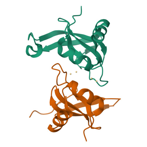
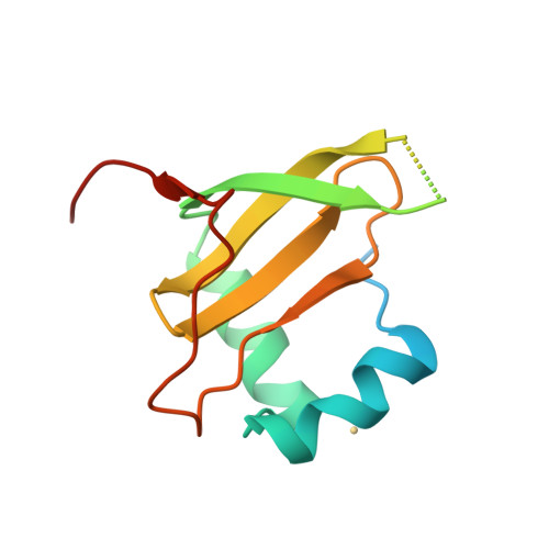
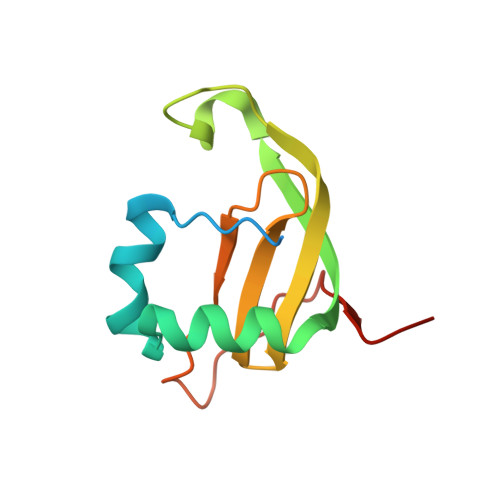
Colicin E5 cleaves tRNAs for Tyr, His, Asn and Asp in their anticodons to abolish protein synthesis in Escherichia coli. We previously showed how its C-terminal RNase domain, E5-CRD, recognizes the anticodon bases but the catalytic mechanism remained to be elucidated. Although the reaction products with 5'-OH and 2',3'-cyclic phosphate ends suggested a similar mechanism to those of RNases A and T1, E5-CRD does not have the His residues necessary as a catalyst in usual RNases. To identify residues important for the catalytic reaction, mutants as to all residues within 5 Å from the central phosphorus of the scissile phosphodiester bond were prepared. Evaluation of the killing activities of the mutant colicins and the RNase activities of the mutant E5-CRDs suggested direct involvement of Arg33, Lys25, Gln29 and Lys60 in the reaction. Particularly, Arg33 plays a critical role and Ile94 provides a structural support of Arg33. Crystal structure of the complex of E5-CRD(R33Q)/dGpdUp showed structural and binding functional integrity of this mutant protein, suggesting involvement of Arg33 in the catalytic reaction. The structure of the free E5-CRD, we also determined, showed great flexibility of a flap region, which facilitates the access of Lys60 to the substrate in an induced-fit manner.
Organizational Affiliation:
Department of Biotechnology, The University of Tokyo, Yayoi 1-1-1, Tokyo 113-8654, Japan.


