3BO5 | pdb_00003bo5

Crystal structure of methyltransferase domain of human Histone-lysine N-methyltransferase SETMAR


Experimental Data Snapshot

  • Method: X-RAY DIFFRACTION
  • Resolution: 1.59 Å
  • R-Value Free: 
    0.199 (Depositor), 0.200 (DCC) 
  • R-Value Work: 
    0.155 (Depositor), 0.150 (DCC) 
  • R-Value Observed: 
    0.157 (Depositor) 

wwPDB Validation   3D Report Full Report


Ligand Structure Quality Assessment 

Created with Raphaël 2.3.0Worse 01 BetterLigand structure goodness of fit to experimental dataBest fitted SAHClick on this verticalbar to view details

This is version 1.3 of the entry. See complete history


Literature

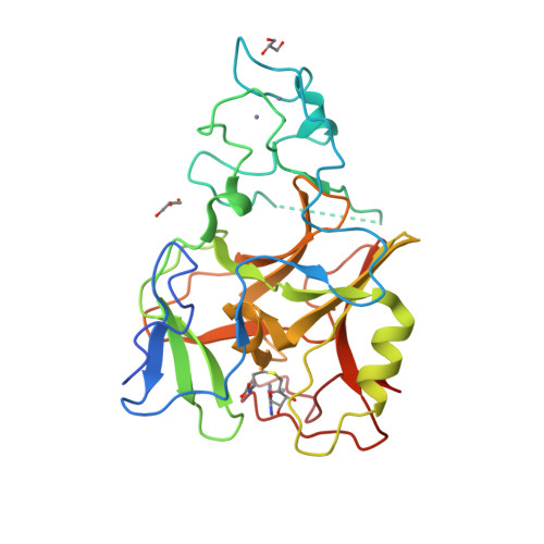
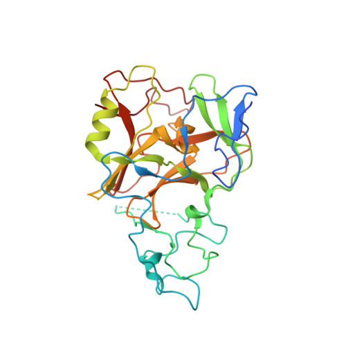
The Crystal Structure of Methyltransferase Domain of Human Histone-lysine N-methyltransferase SETMAR in Complex With AdoHcy.

Wu, H.Lunin, V.V.Ren, H.Dobrovetsky, E.Weigelt, J.Arrowsmith, C.H.Edwards, A.M.Bochkarev, A.Min, J.Plotnikov, A.N.

To be published.

Macromolecules
Find similar proteins by:  (by identity cutoff)  |  3D Structure
Entity ID: 1
MoleculeChains Sequence LengthOrganismDetailsImage
Histone-lysine N-methyltransferase SETMAR290Homo sapiensMutation(s): 2 
Gene Names: SETMAR
EC: 2.1.1.43 (PDB Primary Data), 3.1 (UniProt), 2.1.1.357 (UniProt)
UniProt & NIH Common Fund Data Resources
Find proteins for Q53H47 (Homo sapiens)
Explore Q53H47 
Go to UniProtKB:  Q53H47
PHAROS:  Q53H47
GTEx:  ENSG00000170364 
Entity Groups  
Sequence Clusters30% Identity50% Identity70% Identity90% Identity95% Identity100% Identity
UniProt GroupQ53H47
Sequence Annotations
Expand
  • Reference Sequence
Small Molecules
Ligands 3 Unique
IDChains Name / Formula / InChI Key2D Diagram3D Interactions
SAH
Query on SAH

Download Ideal Coordinates CCD File 
F [auth A]S-ADENOSYL-L-HOMOCYSTEINE
C14 H20 N6 O5 S
ZJUKTBDSGOFHSH-WFMPWKQPSA-N
GOL
Query on GOL

Download Ideal Coordinates CCD File 
G [auth A],
H [auth A]
GLYCEROL
C3 H8 O3
PEDCQBHIVMGVHV-UHFFFAOYSA-N
ZN
Query on ZN

Download Ideal Coordinates CCD File 
B [auth A],
C [auth A],
D [auth A],
E [auth A]
ZINC ION
Zn
PTFCDOFLOPIGGS-UHFFFAOYSA-N
Experimental Data & Validation

Experimental Data

  • Method: X-RAY DIFFRACTION
  • Resolution: 1.59 Å
  • R-Value Free:  0.199 (Depositor), 0.200 (DCC) 
  • R-Value Work:  0.155 (Depositor), 0.150 (DCC) 
  • R-Value Observed: 0.157 (Depositor) 
Space Group: P 1 21 1
Unit Cell:
Length ( Å )Angle ( ˚ )
a = 41.396α = 90
b = 67.827β = 105.93
c = 44.637γ = 90
Software Package:
Software NamePurpose
REFMACrefinement
ADSCdata collection
HKL-3000data reduction
HKL-3000data scaling
SHELXDphasing

Structure Validation

View Full Validation Report


Ligand Structure Quality Assessment 

Created with Raphaël 2.3.0Worse 01 BetterLigand structure goodness of fit to experimental dataBest fitted SAHClick on this verticalbar to view details

Entry History 

Revision History  (Full details and data files)

  • Version 1.0: 2008-01-22
    Type: Initial release
  • Version 1.1: 2011-07-13
    Changes: Non-polymer description, Version format compliance
  • Version 1.2: 2021-10-20
    Changes: Database references, Derived calculations
  • Version 1.3: 2024-02-21
    Changes: Data collection