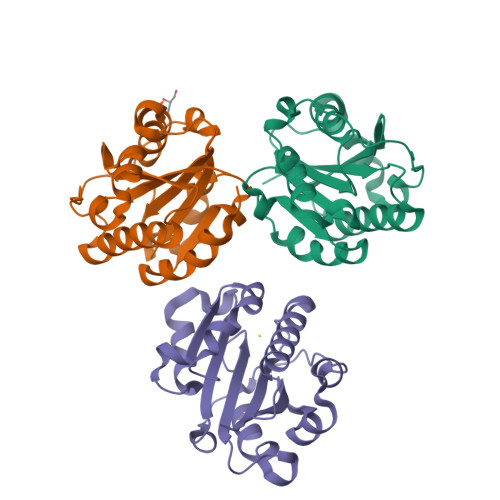
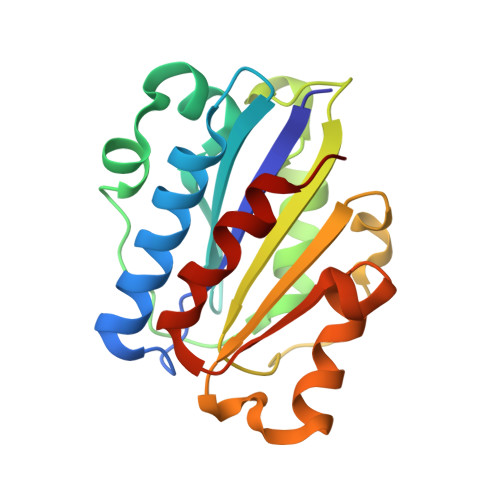
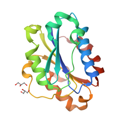
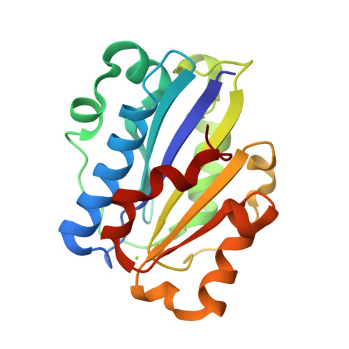
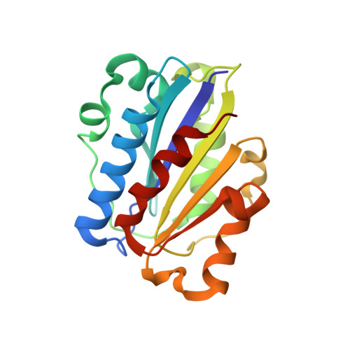
Crystal structure of isoflurane bound to integrin LFA-1 supports a unified mechanism of volatile anesthetic action in the immune and central nervous systems.
Zhang, H., Astrof, N.S., Liu, J.H., Wang, J.H., Shimaoka, M.(2009) FASEB J 23: 2735-2740
- PubMed: 19332643
- DOI: https://doi.org/10.1096/fj.09-129908
- Primary Citation of Related Structures:
3F74, 3F78 - PubMed Abstract:
Volatile anesthetics (VAs), such as isoflurane, induce a general anesthetic state by binding to specific targets (i.e., ion channels) in the central nervous system (CNS). Simultaneously, VAs modulate immune functions, possibly via direct interaction with alternative targets on leukocytes. One such target, the integrin lymphocyte function-associated antigen-1 (LFA-1), has been shown previously to be inhibited by isoflurane. A better understanding of the mechanism by which isoflurane alters protein function requires the detailed information about the drug-protein interaction at an atomic level. Here, we describe the crystal structure of the LFA-1 ligand-binding domain (I domain) in complex with isoflurane at 1.6 A. We discovered that isoflurane binds to an allosteric cavity previously implicated as critical for the transition of LFA-1 from the low- to the high-affinity state. The isoflurane binding site in the I domain involves an array of amphiphilic interactions, thereby resembling a "common anesthetic binding motif" previously predicted for authentic VA binding sites. These results suggest that the allosteric modulation of protein function by isoflurane, as demonstrated for the integrin LFA-1, might represent a unified mechanism shared by the interactions of volatile anesthetics with targets in the CNS.
Organizational Affiliation:
Dana-Farber Cancer Institute, Boston, Massachusetts, USA.


