3H0Y | pdb_00003h0y

Aurora A in complex with a bisanilinopyrimidine


Experimental Data Snapshot

  • Method: X-RAY DIFFRACTION
  • Resolution: 2.50 Å
  • R-Value Free: 
    0.285 (Depositor), 0.280 (DCC) 
  • R-Value Work: 
    0.208 (Depositor), 0.210 (DCC) 
  • R-Value Observed: 
    0.216 (Depositor) 

wwPDB Validation   3D Report Full Report


Ligand Structure Quality Assessment 

Created with Raphaël 2.3.0Worse 01 BetterLigand structure goodness of fit to experimental dataBest fitted 48BClick on this verticalbar to view details

This is version 1.3 of the entry. See complete history


Literature

A class of 2,4-bisanilinopyrimidine Aurora A inhibitors with unusually high selectivity against Aurora B.

Aliagas-Martin, I.Burdick, D.Corson, L.Dotson, J.Drummond, J.Fields, C.Huang, O.W.Hunsaker, T.Kleinheinz, T.Krueger, E.Liang, J.Moffat, J.Phillips, G.Pulk, R.Rawson, T.E.Ultsch, M.Walker, L.Wiesmann, C.Zhang, B.Zhu, B.Y.Cochran, A.G.

(2009) J Med Chem 52: 3300-3307

  • DOI: https://doi.org/10.1021/jm9000314
  • Primary Citation of Related Structures:  
    3H0Y, 3H0Z, 3H10

  • PubMed Abstract: 

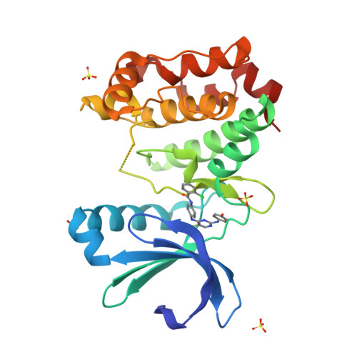
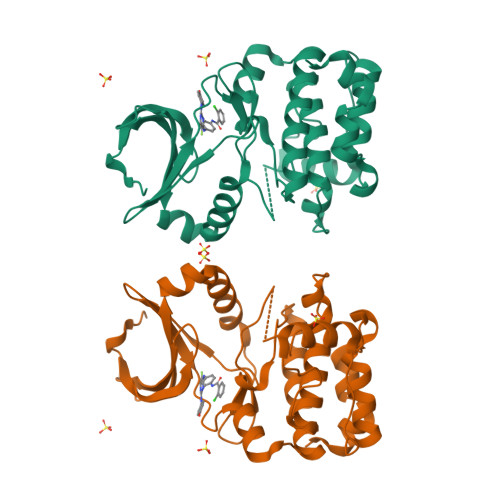
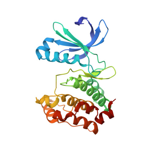
    The two major Aurora kinases carry out critical functions at distinct mitotic stages. Selective inhibitors of these kinases, as well as pan-Aurora inhibitors, show antitumor efficacy and are now under clinical investigation. However, the ATP-binding sites of Aurora A and Aurora B are virtually identical, and the structural basis for selective inhibition has therefore not been clear. We report here a class of bisanilinopyrimidine Aurora A inhibitors with excellent selectivity for Aurora A over Aurora B, both in enzymatic assays and in cellular phenotypic assays. Crystal structures of two of the inhibitors in complex with Aurora A implicate a single amino acid difference in Aurora B as responsible for poor inhibitory activity against this enzyme. Mutation of this residue in Aurora B (E161T) or Aurora A (T217E) is sufficient to swap the inhibition profile, suggesting that this difference might be exploited more generally to achieve high selectivity for Aurora A.


  • Organizational Affiliation

    Department of Small Molecule Drug Discovery, Genentech, Inc., 1 DNA Way, South San Francisco, California 94080, USA.


Macromolecules
Find similar proteins by:  (by identity cutoff)  |  3D Structure
Entity ID: 1
MoleculeChains Sequence LengthOrganismDetailsImage
Serine/threonine-protein kinase 6268Homo sapiensMutation(s): 3 
Gene Names: AURKAAIKARK1AURABTAKSTK15STK6
EC: 2.7.11.1
UniProt & NIH Common Fund Data Resources
Find proteins for O14965 (Homo sapiens)
Explore O14965 
Go to UniProtKB:  O14965
PHAROS:  O14965
GTEx:  ENSG00000087586 
Entity Groups  
Sequence Clusters30% Identity50% Identity70% Identity90% Identity95% Identity100% Identity
UniProt GroupO14965
Sequence Annotations
Expand
  • Reference Sequence
Experimental Data & Validation

Experimental Data

  • Method: X-RAY DIFFRACTION
  • Resolution: 2.50 Å
  • R-Value Free:  0.285 (Depositor), 0.280 (DCC) 
  • R-Value Work:  0.208 (Depositor), 0.210 (DCC) 
  • R-Value Observed: 0.216 (Depositor) 
Space Group: P 61 2 2
Unit Cell:
Length ( Å )Angle ( ˚ )
a = 81.377α = 90
b = 81.377β = 90
c = 170.892γ = 120
Software Package:
Software NamePurpose
HKL-2000data collection
AMoREphasing
REFMACrefinement
DENZOdata reduction
SCALEPACKdata scaling

Structure Validation

View Full Validation Report


Ligand Structure Quality Assessment 

Created with Raphaël 2.3.0Worse 01 BetterLigand structure goodness of fit to experimental dataBest fitted 48BClick on this verticalbar to view details

Entry History 

Deposition Data

Revision History  (Full details and data files)

  • Version 1.0: 2009-07-07
    Type: Initial release
  • Version 1.1: 2011-07-13
    Changes: Advisory, Refinement description, Version format compliance
  • Version 1.2: 2021-10-13
    Changes: Database references, Derived calculations
  • Version 1.3: 2024-02-21
    Changes: Data collection