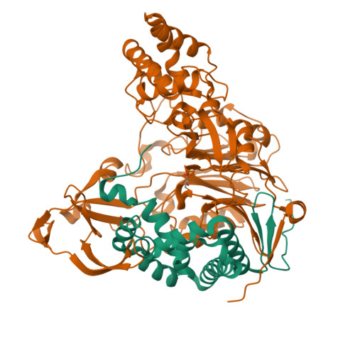
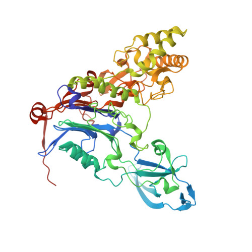
Structural features of cephalosporin acylase reveal the basis of autocatalytic activation.
Cho, K.J., Kim, J.K., Lee, J.H., Shin, H.J., Park, S.S., Kim, K.H.(2009) Biochem Biophys Res Commun 390: 342-348
- PubMed: 19800869
- DOI: https://doi.org/10.1016/j.bbrc.2009.09.134
- Primary Citation of Related Structures:
3JTQ, 3JTR - PubMed Abstract:
Cephalosporin acylase (CA), a member of the N-terminal nucleophile hydrolase family, is activated through two steps of intramolecular autoproteolysis, the first mediated by a serine residue, and the second by a glutamate, which releases the pro-segment and produces an active enzyme. In this study, we have determined the crystal structures of mutants which could affect primary or secondary auto-cleavage and of sequential intermediates of a slow-processing mutant at 2.0-2.5A resolutions. The pro-segments of the mutants undergo dynamic conformational changes during activation and adopt surprisingly different loop conformations from one another. However, the autoproteolytic site was found to form a catalytically competent conformation with a solvent water molecule, which was essentially conserved in the CA mutants.
Organizational Affiliation:
Department of Life Sciences and Biotechnology, Korea University, Seoul 136-701, Republic of Korea.