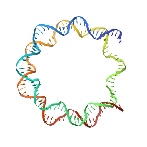
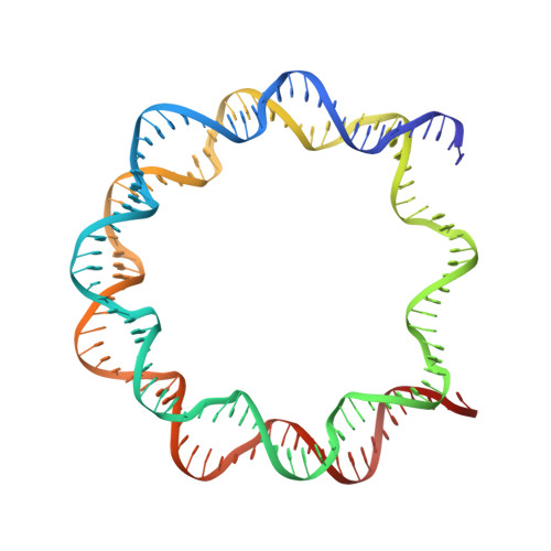
3LJA

Using Soft X-Rays for a Detailed Picture of Divalent Metal Binding in the Nucleosome


Experimental Data Snapshot

  • Method: X-RAY DIFFRACTION
  • Resolution: 2.75 Å
  • R-Value Free: 0.259 
  • R-Value Work: 0.223 
  • R-Value Observed: 0.223 

Starting Model: experimental
View more details

wwPDB Validation   3D Report Full Report


This is version 1.3 of the entry. See complete history


Literature

Using soft X-rays for a detailed picture of divalent metal binding in the nucleosome

Wu, B.Davey, C.A.

(2010) J Mol Biol 398: 633-640

  • DOI: https://doi.org/10.1016/j.jmb.2010.03.038
  • Primary Citation of Related Structures:  
    3LJA

  • PubMed Abstract: 

    Divalent metals associate with DNA in a site-selective manner, which can influence nucleosome positioning, mobility, compaction, and recognition by nuclear factors. We previously characterized divalent metal binding in the nucleosome core using hard (short-wavelength) X-rays allowing high-resolution crystallographic determination of the strongest affinity sites, which revealed that Mn(2+) associates with the DNA major groove in a sequence- and conformation-dependent manner. In this study, we obtained diffraction data with soft X-rays at the Mn(2+) absorption edge for a core particle crystal in the presence of 10 mM MnSO(4), mimicking prevailing Mg(2+) concentration in the nucleus. This provides an exceptional view of counterion binding in the nucleosome through identification of 45 divalent metal binding sites. In addition to that at the well-characterized major interparticle interface, only one other histone-divalent metal binding site is found, which corresponds to a symmetry-related counterpart on the 'free' H2B alpha1 helix C-terminus. This emphasizes the importance of the alpha-helix dipole in ion binding and suggests that the H2B motif may serve as a nucleation site in nucleosome compaction. The 43 sites associated with the DNA are characterized by (1) high-affinity direct coordination at the most electrostatically favorable major groove locations, (2) metal hydrate binding to the major groove, (3) direct coordination to phosphate groups at sites of high charge density, (4) metal hydrate binding in the minor groove, or (5) metal hydrate-divalent anion pairing. Metal hydrates are found within the minor groove only at locations displaying a narrow range of high-intermediate width and to which histone N-terminal tails are not associated or proximal. This indicates that divalent metals and histone tails can both collaborate and compete in minor groove association, which sheds light on nucleosome solubility and chromatin compaction behavior.


  • Organizational Affiliation

    Division of Structural and Computational Biology, School of Biological Sciences, Nanyang Technological University, 60 Nanyang Drive, Singapore 637551, Singapore.


Macromolecules

Find similar proteins by:  (by identity cutoff)  |  3D Structure
Entity ID: 1
MoleculeChains Sequence LengthOrganismDetailsImage
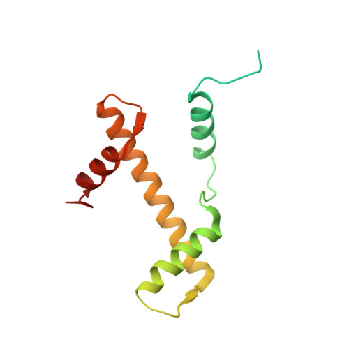
Histone H3.2
A, E
135Xenopus laevisMutation(s): 0 
UniProt
Find proteins for P84233 (Xenopus laevis)
Explore P84233 
Go to UniProtKB:  P84233
Entity Groups  
Sequence Clusters30% Identity50% Identity70% Identity90% Identity95% Identity100% Identity
UniProt GroupP84233
Sequence Annotations
Expand
  • Reference Sequence
Find similar proteins by:  (by identity cutoff)  |  3D Structure
Entity ID: 2
MoleculeChains Sequence LengthOrganismDetailsImage
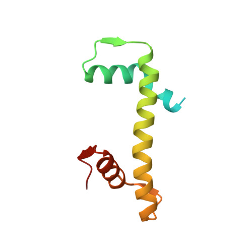
Histone H4
B, F
102Xenopus laevisMutation(s): 0 
UniProt
Find proteins for P62799 (Xenopus laevis)
Explore P62799 
Go to UniProtKB:  P62799
Entity Groups  
Sequence Clusters30% Identity50% Identity70% Identity90% Identity95% Identity100% Identity
UniProt GroupP62799
Sequence Annotations
Expand
  • Reference Sequence
Find similar proteins by:  (by identity cutoff)  |  3D Structure
Entity ID: 3
MoleculeChains Sequence LengthOrganismDetailsImage
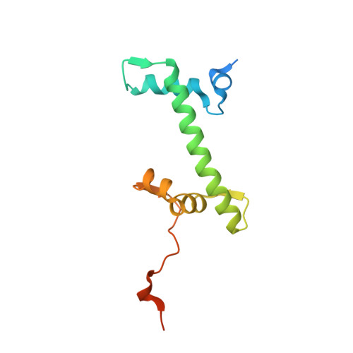
Histone H2A
C, G
129Xenopus laevisMutation(s): 0 
Gene Names: LOC494591
UniProt
Find proteins for P06897 (Xenopus laevis)
Explore P06897 
Go to UniProtKB:  P06897
Entity Groups  
Sequence Clusters30% Identity50% Identity70% Identity90% Identity95% Identity100% Identity
UniProt GroupP06897
Sequence Annotations
Expand
  • Reference Sequence
Find similar proteins by:  (by identity cutoff)  |  3D Structure
Entity ID: 4
MoleculeChains Sequence LengthOrganismDetailsImage
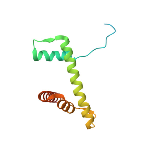
Histone H2B 1.1
D, H
122Xenopus laevisMutation(s): 0 
UniProt
Find proteins for P02281 (Xenopus laevis)
Explore P02281 
Go to UniProtKB:  P02281
Entity Groups  
Sequence Clusters30% Identity50% Identity70% Identity90% Identity95% Identity100% Identity
UniProt GroupP02281
Sequence Annotations
Expand
  • Reference Sequence
Find similar nucleic acids by:  (by identity cutoff)  |  3D Structure
Entity ID: 5
MoleculeChains LengthOrganismImage
147mer DNA147N/A
Sequence Annotations
Expand
  • Reference Sequence
Find similar nucleic acids by:  (by identity cutoff)  |  3D Structure
Entity ID: 6
MoleculeChains LengthOrganismImage
147mer DNA147N/A
Sequence Annotations
Expand
  • Reference Sequence
Small Molecules
Ligands 2 Unique
IDChains Name / Formula / InChI Key2D Diagram3D Interactions
SO4
Query on SO4

Download Ideal Coordinates CCD File 
K [auth D],
M [auth G],
O [auth H]
SULFATE ION
O4 S
QAOWNCQODCNURD-UHFFFAOYSA-L
MN
Query on MN

Download Ideal Coordinates CCD File 
AA [auth I]
AB [auth J]
BA [auth I]
BB [auth J]
CA [auth I]
AA [auth I],
AB [auth J],
BA [auth I],
BB [auth J],
CA [auth I],
CB [auth J],
DA [auth I],
DB [auth J],
EA [auth I],
EB [auth J],
FA [auth I],
FB [auth J],
GA [auth I],
HA [auth J],
IA [auth J],
JA [auth J],
KA [auth J],
L [auth E],
LA [auth J],
MA [auth J],
N [auth H],
NA [auth J],
OA [auth J],
P [auth I],
PA [auth J],
Q [auth I],
QA [auth J],
R [auth I],
RA [auth J],
S [auth I],
SA [auth J],
T [auth I],
TA [auth J],
U [auth I],
UA [auth J],
V [auth I],
VA [auth J],
W [auth I],
WA [auth J],
X [auth I],
XA [auth J],
Y [auth I],
YA [auth J],
Z [auth I],
ZA [auth J]
MANGANESE (II) ION
Mn
WAEMQWOKJMHJLA-UHFFFAOYSA-N
Experimental Data & Validation

Experimental Data

  • Method: X-RAY DIFFRACTION
  • Resolution: 2.75 Å
  • R-Value Free: 0.259 
  • R-Value Work: 0.223 
  • R-Value Observed: 0.223 
  • Space Group: P 21 21 21
Unit Cell:
Length ( Å )Angle ( ˚ )
a = 106.348α = 90
b = 109.785β = 90
c = 182.424γ = 90
Software Package:
Software NamePurpose
MAR345dtbdata collection
AMoREphasing
REFMACrefinement
MOSFLMdata reduction
SCALAdata scaling

Structure Validation

View Full Validation Report


Entry History 

Deposition Data

Revision History  (Full details and data files)

  • Version 1.0: 2010-04-14
    Type: Initial release
  • Version 1.1: 2011-07-13
    Changes: Version format compliance
  • Version 1.2: 2014-02-12
    Changes: Database references
  • Version 1.3: 2023-11-01
    Changes: Data collection, Database references, Derived calculations, Refinement description