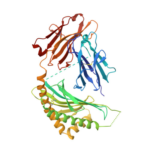
3OV6

CD1c in complex with MPM (mannosyl-beta1-phosphomycoketide)


Experimental Data Snapshot

  • Method: X-RAY DIFFRACTION
  • Resolution: 2.50 Å
  • R-Value Free: 0.273 
  • R-Value Work: 0.240 
  • R-Value Observed: 0.242 

wwPDB Validation   3D Report Full Report


Ligand Structure Quality Assessment 


This is version 1.4 of the entry. See complete history


Literature

The 2.5 A structure of CD1c in complex with a mycobacterial lipid reveals an open groove ideally suited for diverse antigen presentation

Scharf, L.Li, N.S.Hawk, A.J.Garzon, D.Zhang, T.Fox, L.M.Kazen, A.R.Shah, S.Haddadian, E.J.Gumperz, J.E.Saghatelian, A.Faraldo-Gomez, J.D.Meredith, S.C.Piccirilli, J.A.Adams, E.J.

(2010) Immunity 33: 853-862

  • DOI: https://doi.org/10.1016/j.immuni.2010.11.026
  • Primary Citation of Related Structures:  
    3OV6

  • PubMed Abstract: 

    CD1 molecules function to present lipid-based antigens to T cells. Here we present the crystal structure of CD1c at 2.5 Å resolution, in complex with the pathogenic Mycobacterium tuberculosis antigen mannosyl-β1-phosphomycoketide (MPM). CD1c accommodated MPM's methylated alkyl chain exclusively in the A' pocket, aided by a unique exit portal underneath the α1 helix. Most striking was an open F' pocket architecture lacking the closed cavity structure of other CD1 molecules, reminiscent of peptide binding grooves of classical major histocompatibility complex molecules. This feature, combined with tryptophan-fluorescence quenching during loading of a dodecameric lipopeptide antigen, provides a compelling model by which both the lipid and peptide moieties of the lipopeptide are involved in CD1c presentation of lipopeptides.


  • Organizational Affiliation

    Department of Biochemistry and Molecular Biology, University of Chicago, Chicago, IL 60637, USA.


Macromolecules
Find similar proteins by:  (by identity cutoff)  |  3D Structure
Entity ID: 1
MoleculeChains Sequence LengthOrganismDetailsImage
Beta-2-microglobulin, T-cell surface glycoprotein CD1c, T-cell surface glycoprotein CD1b397Homo sapiensMutation(s): 6 
UniProt & NIH Common Fund Data Resources
Find proteins for P29016 (Homo sapiens)
Explore P29016 
Go to UniProtKB:  P29016
PHAROS:  P29016
GTEx:  ENSG00000158485 
Find proteins for P29017 (Homo sapiens)
Explore P29017 
Go to UniProtKB:  P29017
PHAROS:  P29017
GTEx:  ENSG00000158481 
Find proteins for P61769 (Homo sapiens)
Explore P61769 
Go to UniProtKB:  P61769
PHAROS:  P61769
GTEx:  ENSG00000166710 
Entity Groups  
Sequence Clusters30% Identity50% Identity70% Identity90% Identity95% Identity100% Identity
UniProt GroupsP29017P29016P61769
Glycosylation
Glycosylation Sites: 1Go to GlyGen: P61769-1P29016-1P29017-1
Sequence Annotations
Expand
  • Reference Sequence
Small Molecules
Ligands 3 Unique
IDChains Name / Formula / InChI Key2D Diagram3D Interactions
MK0
Query on MK0

Download Ideal Coordinates CCD File 
D [auth A]1-O-[(S)-hydroxy{[(4S,8S,16S,20S)-4,8,12,16,20-pentamethylheptacosyl]oxy}phosphoryl]-beta-D-mannopyranose
C38 H77 O9 P
BWDAWKXFHWFXEQ-RIEBCBCMSA-N
NAG
Query on NAG

Download Ideal Coordinates CCD File 
B [auth A]2-acetamido-2-deoxy-beta-D-glucopyranose
C8 H15 N O6
OVRNDRQMDRJTHS-FMDGEEDCSA-N
D12
Query on D12

Download Ideal Coordinates CCD File 
C [auth A]DODECANE
C12 H26
SNRUBQQJIBEYMU-UHFFFAOYSA-N
Experimental Data & Validation

Experimental Data

  • Method: X-RAY DIFFRACTION
  • Resolution: 2.50 Å
  • R-Value Free: 0.273 
  • R-Value Work: 0.240 
  • R-Value Observed: 0.242 
  • Space Group: P 21 21 21
Unit Cell:
Length ( Å )Angle ( ˚ )
a = 53.724α = 90
b = 87.087β = 90
c = 88.808γ = 90
Software Package:
Software NamePurpose
HKL-2000data collection
PHASERphasing
PHENIXrefinement
HKL-2000data reduction
HKL-2000data scaling

Structure Validation

View Full Validation Report


Ligand Structure Quality Assessment 


Entry History 

Revision History  (Full details and data files)

  • Version 1.0: 2011-01-19
    Type: Initial release
  • Version 1.1: 2011-07-13
    Changes: Version format compliance
  • Version 1.2: 2017-07-26
    Changes: Advisory, Data collection, Refinement description, Source and taxonomy
  • Version 1.3: 2020-07-29
    Type: Remediation
    Reason: Carbohydrate remediation
    Changes: Data collection, Database references, Derived calculations, Structure summary
  • Version 1.4: 2024-11-20
    Changes: Advisory, Data collection, Database references, Structure summary