RCSB PDB - 3RTI: Crystal structure of ricin bound with formycin monophosphate

 3RTI

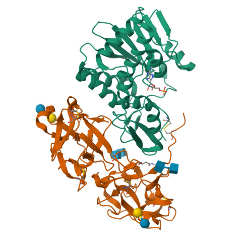
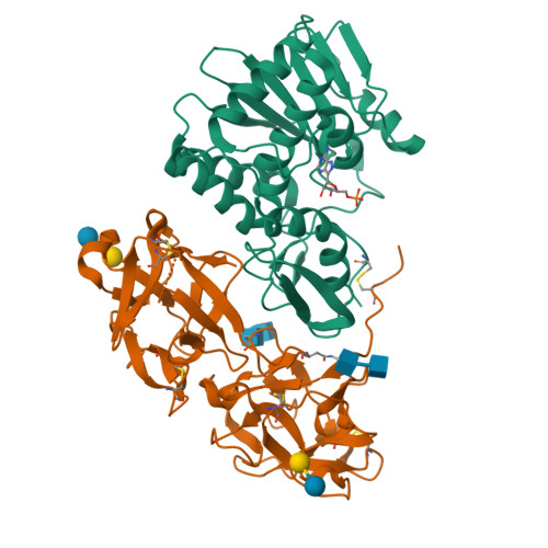
Crystal structure of ricin bound with formycin monophosphate


Experimental Data Snapshot

  • Method: X-RAY DIFFRACTION
  • Resolution: 2.80 Å
  • R-Value Work: 0.209 

wwPDB Validation   3D Report Full Report


Ligand Structure Quality Assessment 

Created with Raphaël 2.3.0Worse 01 BetterLigand structure goodness of fit to experimental dataBest fitted FMPClick on this verticalbar to view details

This is version 2.1 of the entry. See complete history


Literature

X-ray analysis of substrate analogs in the ricin A-chain active site.

Monzingo, A.F.Robertus, J.D.

(1992) J Mol Biol 227: 1136-1145

  • DOI: https://doi.org/10.1016/0022-2836(92)90526-p
  • Primary Citation of Related Structures:  
    3RTI, 3RTJ

  • PubMed Abstract: 

    Ricin A-chain is an N-glycosidase that hydrolyzes the adenine ring from a specific adenosine of rRNA. Formycin monophosphate (FMP) and adenyl(3'-->5')guanosine (ApG) were bound to ricin A-chain and their structures elucidated by X-ray crystallography. The formycin ring stacks between tyrosines 80 and 123 and at least four hydrogen bonds are made to the adenine moiety. A residue invariant in this enzyme class, Arg180, appears to hydrogen bond to N-3 of the susceptible adenine. Three hypothetical models for binding a true hexanucleotide substrate, CGAGAG, are proposed. They incorporate adenine binding, shown by crystallography, but also include geometry likely to favor catalysis. For example, efforts have been made to orient the ribose ring in a way that allows solvent attack and oxycarbonium stabilization by the enzyme. The favored model is a simple perturbation of the tetraloop structure determined by nuclear magnetic resonance for similar polynucleotides. The model is attractive in that specific roles are defined for conserved protein residues. A mechanism of action is proposed. It invokes oxycarbonium ion stabilization on ribose by Glu177 in the transition state. Arg180 stabilizes anion development on the leaving adenine by protonation at N-3 and may activate a trapped water molecule that is the ultimate nucleophile in the depurination.


  • Organizational Affiliation

    Clayton Foundation Biochemical Institute, Department of Chemistry and Biochemistry, University of Texas, Austin 78712.


Macromolecules
Find similar proteins by:  (by identity cutoff)  |  3D Structure
Entity ID: 1
MoleculeChains Sequence LengthOrganismDetailsImage
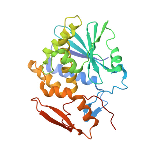
Ricin267Ricinus communisMutation(s): 0 
EC: 3.2.2.22
UniProt
Find proteins for P02879 (Ricinus communis)
Explore P02879 
Go to UniProtKB:  P02879
Entity Groups  
Sequence Clusters30% Identity50% Identity70% Identity90% Identity95% Identity100% Identity
UniProt GroupP02879
Sequence Annotations
Expand
  • Reference Sequence
Find similar proteins by:  (by identity cutoff)  |  3D Structure
Entity ID: 2
MoleculeChains Sequence LengthOrganismDetailsImage
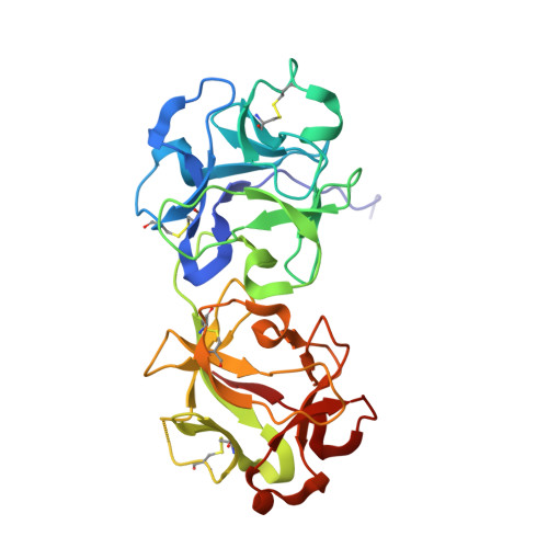
Ricin262Ricinus communisMutation(s): 0 
EC: 3.2.2.22
UniProt
Find proteins for P02879 (Ricinus communis)
Explore P02879 
Go to UniProtKB:  P02879
Entity Groups  
Sequence Clusters30% Identity50% Identity70% Identity90% Identity95% Identity100% Identity
UniProt GroupP02879
Glycosylation
Glycosylation Sites: 2
Sequence Annotations
Expand
  • Reference Sequence
Oligosaccharides

Help

Entity ID: 3
MoleculeChains Length2D Diagram Glycosylation3D Interactions
beta-D-galactopyranose-(1-4)-beta-D-glucopyranose
C, D
2N/A
Glycosylation Resources
GlyTouCan:  G84224TW
GlyCosmos:  G84224TW
GlyGen:  G84224TW
Entity ID: 4
MoleculeChains Length2D Diagram Glycosylation3D Interactions
2-acetamido-2-deoxy-beta-D-glucopyranose-(1-4)-2-acetamido-2-deoxy-beta-D-glucopyranose
E, F
2N-Glycosylation
Glycosylation Resources
GlyTouCan:  G42666HT
GlyCosmos:  G42666HT
GlyGen:  G42666HT
Small Molecules
Ligands 1 Unique
IDChains Name / Formula / InChI Key2D Diagram3D Interactions
FMP
Query on FMP

Download Ideal Coordinates CCD File 
G [auth A]FORMYCIN-5'-MONOPHOSPHATE
C10 H14 N5 O7 P
PBAHXXBYQACZMA-KSYZLYKTSA-N
Binding Affinity Annotations 
IDSourceBinding Affinity
FMP PDBBind:  3RTI Ki: 43 (nM) from 1 assay(s)
Biologically Interesting Molecules (External Reference) 1 Unique
Experimental Data & Validation

Experimental Data

  • Method: X-RAY DIFFRACTION
  • Resolution: 2.80 Å
  • R-Value Work: 0.209 
  • Space Group: P 21 21 21
Unit Cell:
Length ( Å )Angle ( ˚ )
a = 72.74α = 90
b = 78.49β = 90
c = 114.34γ = 90
Software Package:
Software NamePurpose
Sandata collection
X-PLORmodel building
X-PLORrefinement
Sandata reduction
Sandata scaling
X-PLORphasing

Structure Validation

View Full Validation Report


Ligand Structure Quality Assessment 

Created with Raphaël 2.3.0Worse 01 BetterLigand structure goodness of fit to experimental dataBest fitted FMPClick on this verticalbar to view details

Entry History 

Deposition Data

Revision History  (Full details and data files)

  • Version 1.0: 2011-08-31
    Type: Initial release
  • Version 2.0: 2020-07-29
    Type: Remediation
    Reason: Carbohydrate remediation
    Changes: Atomic model, Data collection, Derived calculations, Structure summary
  • Version 2.1: 2024-11-20
    Changes: Data collection, Database references, Structure summary