3S11

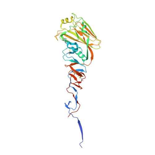
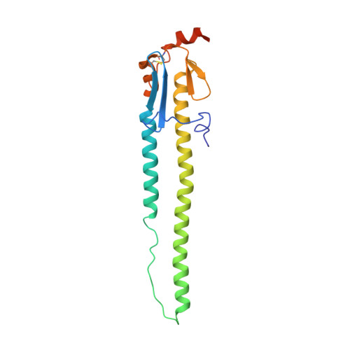
Crystal structure of H5N1 influenza virus hemagglutinin, strain 437-10


Experimental Data Snapshot

  • Method: X-RAY DIFFRACTION
  • Resolution: 2.50 Å
  • R-Value Free: 0.265 
  • R-Value Work: 0.221 
  • R-Value Observed: 0.223 

wwPDB Validation   3D Report Full Report


Ligand Structure Quality Assessment 


This is version 3.0 of the entry. See complete history


Literature

Acid stability of the hemagglutinin protein regulates H5N1 influenza virus pathogenicity.

DuBois, R.M.Zaraket, H.Reddivari, M.Heath, R.J.White, S.W.Russell, C.J.

(2011) PLoS Pathog 7: e1002398-e1002398

  • DOI: https://doi.org/10.1371/journal.ppat.1002398
  • Primary Citation of Related Structures:  
    3S11, 3S12, 3S13

  • PubMed Abstract: 

    Highly pathogenic avian influenza viruses of the H5N1 subtype continue to threaten agriculture and human health. Here, we use biochemistry and x-ray crystallography to reveal how amino-acid variations in the hemagglutinin (HA) protein contribute to the pathogenicity of H5N1 influenza virus in chickens. HA proteins from highly pathogenic (HP) A/chicken/Hong Kong/YU562/2001 and moderately pathogenic (MP) A/goose/Hong Kong/437-10/1999 isolates of H5N1 were found to be expressed and cleaved in similar amounts, and both proteins had similar receptor-binding properties. However, amino-acid variations at positions 104 and 115 in the vestigial esterase sub-domain of the HA1 receptor-binding domain (RBD) were found to modulate the pH of HA activation such that the HP and MP HA proteins are activated for membrane fusion at pH 5.7 and 5.3, respectively. In general, an increase in H5N1 pathogenicity in chickens was found to correlate with an increase in the pH of HA activation for mutant and chimeric HA proteins in the observed range of pH 5.2 to 6.0. We determined a crystal structure of the MP HA protein at 2.50 Å resolution and two structures of HP HA at 2.95 and 3.10 Å resolution. Residues 104 and 115 that modulate the acid stability of the HA protein are situated at the N- and C-termini of the 110-helix in the vestigial esterase sub-domain, which interacts with the B loop of the HA2 stalk domain. Interactions between the 110-helix and the stalk domain appear to be important in regulating HA protein acid stability, which in turn modulates influenza virus replication and pathogenesis. Overall, an optimal activation pH of the HA protein is found to be necessary for high pathogenicity by H5N1 influenza virus in avian species.


  • Organizational Affiliation

    Department of Structural Biology, St. Jude Children's Research Hospital, Memphis, Tennessee United States of America.


Macromolecules
Find similar proteins by:  (by identity cutoff)  |  3D Structure
Entity ID: 1
MoleculeChains Sequence LengthOrganismDetailsImage
Hemagglutinin HA1 chain
A, C, E
331Influenza A virusMutation(s): 0 
UniProt
Find proteins for Q9Q0U6 (Influenza A virus (strain A/Goose/Guangdong/1/1996 H5N1 genotype Gs/Gd))
Explore Q9Q0U6 
Go to UniProtKB:  Q9Q0U6
Entity Groups  
Sequence Clusters30% Identity50% Identity70% Identity90% Identity95% Identity100% Identity
UniProt GroupQ9Q0U6
Glycosylation
Glycosylation Sites: 1
Sequence Annotations
Expand
  • Reference Sequence
Find similar proteins by:  (by identity cutoff)  |  3D Structure
Entity ID: 2
MoleculeChains Sequence LengthOrganismDetailsImage
Hemagglutinin HA2 chain
B, D, F
182Influenza A virusMutation(s): 0 
UniProt
Find proteins for Q8QPL1 (Influenza A virus (strain A/Duck/Hong Kong/2986.1/2000 H5N1 genotype C))
Explore Q8QPL1 
Go to UniProtKB:  Q8QPL1
Entity Groups  
Sequence Clusters30% Identity50% Identity70% Identity90% Identity95% Identity100% Identity
UniProt GroupQ8QPL1
Sequence Annotations
Expand
  • Reference Sequence
Oligosaccharides

Help

Entity ID: 3
MoleculeChains Length2D Diagram Glycosylation3D Interactions
2-acetamido-2-deoxy-beta-D-glucopyranose-(1-4)-2-acetamido-2-deoxy-beta-D-glucopyranose
G, I
2N/A
Glycosylation Resources
GlyTouCan:  G42666HT
GlyCosmos:  G42666HT
GlyGen:  G42666HT
Entity ID: 4
MoleculeChains Length2D Diagram Glycosylation3D Interactions
beta-D-mannopyranose-(1-4)-2-acetamido-2-deoxy-beta-D-glucopyranose-(1-4)-2-acetamido-2-deoxy-beta-D-glucopyranose
H
3N/A
Glycosylation Resources
GlyTouCan:  G15407YE
GlyCosmos:  G15407YE
GlyGen:  G15407YE
Small Molecules
Ligands 3 Unique
IDChains Name / Formula / InChI Key2D Diagram3D Interactions
NAG
Query on NAG

Download Ideal Coordinates CCD File 
J [auth A],
M [auth C],
O [auth E]
2-acetamido-2-deoxy-beta-D-glucopyranose
C8 H15 N O6
OVRNDRQMDRJTHS-FMDGEEDCSA-N
TAM
Query on TAM

Download Ideal Coordinates CCD File 
L [auth A]TRIS(HYDROXYETHYL)AMINOMETHANE
C7 H17 N O3
GKODZWOPPOTFGA-UHFFFAOYSA-N
GOL
Query on GOL

Download Ideal Coordinates CCD File 
K [auth A],
N [auth C],
P [auth E]
GLYCEROL
C3 H8 O3
PEDCQBHIVMGVHV-UHFFFAOYSA-N
Experimental Data & Validation

Experimental Data

  • Method: X-RAY DIFFRACTION
  • Resolution: 2.50 Å
  • R-Value Free: 0.265 
  • R-Value Work: 0.221 
  • R-Value Observed: 0.223 
  • Space Group: P 1 21 1
Unit Cell:
Length ( Å )Angle ( ˚ )
a = 69.365α = 90
b = 241.054β = 116.69
c = 70.061γ = 90
Software Package:
Software NamePurpose
SCALEPACKdata scaling
PHASERphasing
REFMACrefinement
PDB_EXTRACTdata extraction
HKL-2000data collection
HKL-2000data reduction

Structure Validation

View Full Validation Report


Ligand Structure Quality Assessment 


Entry History 

Deposition Data

Revision History  (Full details and data files)

  • Version 1.0: 2011-12-14
    Type: Initial release
  • Version 1.1: 2012-03-28
    Changes: Database references
  • Version 2.0: 2020-07-29
    Type: Remediation
    Reason: Carbohydrate remediation
    Changes: Atomic model, Data collection, Database references, Derived calculations, Structure summary
  • Version 3.0: 2024-12-25
    Changes: Advisory, Atomic model, Data collection, Database references, Derived calculations, Structure summary