3UBL

Crystal structure of glutathione transferase (TARGET EFI-501770) from leptospira interrogans with gsh bound


Experimental Data Snapshot

  • Method: X-RAY DIFFRACTION
  • Resolution: 2.00 Å
  • R-Value Free: 
    0.235 (Depositor), 0.240 (DCC) 
  • R-Value Work: 
    0.200 (Depositor), 0.200 (DCC) 
  • R-Value Observed: 
    0.201 (Depositor) 

Starting Model: experimental
View more details

wwPDB Validation   3D Report Full Report


Ligand Structure Quality Assessment 

Created with Raphaël 2.3.0Worse 01 BetterLigand structure goodness of fit to experimental dataBest fitted GSHClick on this verticalbar to view details

This is version 1.4 of the entry. See complete history


Literature

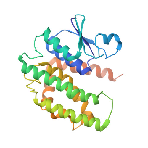
Crystal Structure of Glutathione S-Transferase from Leptospira Interrogans

Patskovsky, Y.Toro, R.Bhosle, R.Zencheck, W.D.Hillerich, B.Seidel, R.D.Washington, E.Scott Glenn, A.Chowdhury, S.Evans, B.Hammonds, J.Imker, H.J.Armstrong, R.N.Gerlt, J.A.Almo, S.C.

To be published.

Macromolecules
Find similar proteins by:  (by identity cutoff)  |  3D Structure
Entity ID: 1
MoleculeChains Sequence LengthOrganismDetailsImage
Glutathione transferase
A, B
242Leptospira interrogans serovar Lai str. 56601Mutation(s): 0 
Gene Names: gstLA_2623
UniProt
Find proteins for Q8F2Y8 (Leptospira interrogans serogroup Icterohaemorrhagiae serovar Lai (strain 56601))
Explore Q8F2Y8 
Go to UniProtKB:  Q8F2Y8
Entity Groups  
Sequence Clusters30% Identity50% Identity70% Identity90% Identity95% Identity100% Identity
UniProt GroupQ8F2Y8
Sequence Annotations
Expand
  • Reference Sequence
Small Molecules
Ligands 5 Unique
IDChains Name / Formula / InChI Key2D Diagram3D Interactions
GSH
Query on GSH

Download Ideal Coordinates CCD File 
C [auth A],
L [auth B]
GLUTATHIONE
C10 H17 N3 O6 S
RWSXRVCMGQZWBV-WDSKDSINSA-N
SO4
Query on SO4

Download Ideal Coordinates CCD File 
D [auth A],
E [auth A],
F [auth A],
M [auth B]
SULFATE ION
O4 S
QAOWNCQODCNURD-UHFFFAOYSA-L
GOL
Query on GOL

Download Ideal Coordinates CCD File 
G [auth A],
I [auth A],
J [auth A],
N [auth B]
GLYCEROL
C3 H8 O3
PEDCQBHIVMGVHV-UHFFFAOYSA-N
CL
Query on CL

Download Ideal Coordinates CCD File 
H [auth A],
O [auth B]
CHLORIDE ION
Cl
VEXZGXHMUGYJMC-UHFFFAOYSA-M
UNX
Query on UNX

Download Ideal Coordinates CCD File 
K [auth A],
P [auth B]
UNKNOWN ATOM OR ION
X
Experimental Data & Validation

Experimental Data

  • Method: X-RAY DIFFRACTION
  • Resolution: 2.00 Å
  • R-Value Free:  0.235 (Depositor), 0.240 (DCC) 
  • R-Value Work:  0.200 (Depositor), 0.200 (DCC) 
  • R-Value Observed: 0.201 (Depositor) 
Space Group: P 43 2 2
Unit Cell:
Length ( Å )Angle ( ˚ )
a = 82.591α = 90
b = 82.591β = 90
c = 175.701γ = 90
Software Package:
Software NamePurpose
PHASERphasing
REFMACrefinement
HKL-2000data reduction
HKL-2000data scaling

Structure Validation

View Full Validation Report


Ligand Structure Quality Assessment 

Created with Raphaël 2.3.0Worse 01 BetterLigand structure goodness of fit to experimental dataBest fitted GSHClick on this verticalbar to view details

Entry History 

Revision History  (Full details and data files)

  • Version 1.0: 2011-11-09
    Type: Initial release
  • Version 1.1: 2011-12-21
    Changes: Non-polymer description
  • Version 1.2: 2012-02-29
    Changes: Database references, Structure summary
  • Version 1.3: 2018-01-24
    Changes: Structure summary
  • Version 1.4: 2023-09-13
    Changes: Data collection, Database references, Derived calculations, Refinement description