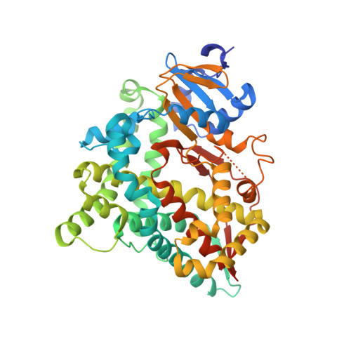
4FDH

Structure of human aldosterone synthase, CYP11B2, in complex with fadrozole


Experimental Data Snapshot

  • Method: X-RAY DIFFRACTION
  • Resolution: 2.71 Å
  • R-Value Free: 0.300 
  • R-Value Work: 0.224 
  • R-Value Observed: 0.228 

wwPDB Validation   3D Report Full Report


Ligand Structure Quality Assessment 


This is version 1.2 of the entry. See complete history


Literature

Structural insights into aldosterone synthase substrate specificity and targeted inhibition.

Strushkevich, N.Gilep, A.A.Shen, L.Arrowsmith, C.H.Edwards, A.M.Usanov, S.A.Park, H.W.

(2013) Mol Endocrinol 27: 315-324

  • DOI: https://doi.org/10.1210/me.2012-1287
  • Primary Citation of Related Structures:  
    4DVQ, 4FDH

  • PubMed Abstract: 

    Aldosterone is a major mineralocorticoid hormone that plays a key role in the regulation of electrolyte balance and blood pressure. Excess aldosterone levels can arise from dysregulation of the renin-angiotensin-aldosterone system and are implicated in the pathogenesis of hypertension and heart failure. Aldosterone synthase (cytochrome P450 11B2, CYP11B2) is the sole enzyme responsible for the production of aldosterone in humans. Blocking of aldosterone synthesis by mediating aldosterone synthase activity is thus a recently emerging pharmacological therapy for hypertension, yet a lack of structural information has limited this approach. Here, we present the crystal structures of human aldosterone synthase in complex with a substrate deoxycorticosterone and an inhibitor fadrozole. The structures reveal a hydrophobic cavity with specific features associated with corticosteroid recognition. The substrate binding mode, along with biochemical data, explains the high 11β-hydroxylase activity of aldosterone synthase toward both gluco- and mineralocorticoid formation. The low processivity of aldosterone synthase with a high extent of intermediates release might be one of the mechanisms of controlled aldosterone production from deoxycorticosterone. Although the active site pocket is lined by identical residues between CYP11B isoforms, most of the divergent residues that confer additional 18-oxidase activity of aldosterone synthase are located in the I-helix (vicinity of the O(2) activation path) and loops around the H-helix (affecting an egress channel closure required for retaining intermediates in the active site). This intrinsic flexibility is also reflected in isoform-selective inhibitor binding. Fadrozole binds to aldosterone synthase in the R-configuration, using part of the active site cavity pointing toward the egress channel. The structural organization of aldosterone synthase provides critical insights into the molecular mechanism of catalysis and enables rational design of more specific antihypertensive agents.


  • Organizational Affiliation

    Structural Genomics Consortium, University of Toronto, Toronto, Ontario, Canada M5G 1L7. [email protected]


Macromolecules
Find similar proteins by:  (by identity cutoff)  |  3D Structure
Entity ID: 1
MoleculeChains Sequence LengthOrganismDetailsImage
Cytochrome P450 11B2, mitochondrial
A, B, C, D, E
A, B, C, D, E, F, G, H, I, J, K, L
483Homo sapiensMutation(s): 0 
Gene Names: Aldosterone synthaseCYP11B2
EC: 1.14.15.4 (PDB Primary Data), 1.14.15.5 (PDB Primary Data)
UniProt & NIH Common Fund Data Resources
Find proteins for P19099 (Homo sapiens)
Explore P19099 
Go to UniProtKB:  P19099
PHAROS:  P19099
GTEx:  ENSG00000179142 
Entity Groups  
Sequence Clusters30% Identity50% Identity70% Identity90% Identity95% Identity100% Identity
UniProt GroupP19099
Sequence Annotations
Expand
  • Reference Sequence
Small Molecules
Ligands 2 Unique
IDChains Name / Formula / InChI Key2D Diagram3D Interactions
HEM
Query on HEM

Download Ideal Coordinates CCD File 
AA [auth H]
CA [auth I]
EA [auth J]
GA [auth K]
IA [auth L]
AA [auth H],
CA [auth I],
EA [auth J],
GA [auth K],
IA [auth L],
M [auth A],
O [auth B],
Q [auth C],
S [auth D],
U [auth E],
W [auth F],
Y [auth G]
PROTOPORPHYRIN IX CONTAINING FE
C34 H32 Fe N4 O4
KABFMIBPWCXCRK-RGGAHWMASA-L
0T3
Query on 0T3

Download Ideal Coordinates CCD File 
BA [auth H]
DA [auth I]
FA [auth J]
HA [auth K]
JA [auth L]
BA [auth H],
DA [auth I],
FA [auth J],
HA [auth K],
JA [auth L],
N [auth A],
P [auth B],
R [auth C],
T [auth D],
V [auth E],
X [auth F],
Z [auth G]
4-[(5R)-5,6,7,8-tetrahydroimidazo[1,5-a]pyridin-5-yl]benzonitrile
C14 H13 N3
CLPFFLWZZBQMAO-CQSZACIVSA-N
Binding Affinity Annotations 
IDSourceBinding Affinity
0T3 BindingDB:  4FDH IC50: min: 0.8, max: 119 (nM) from 17 assay(s)
Experimental Data & Validation

Experimental Data

  • Method: X-RAY DIFFRACTION
  • Resolution: 2.71 Å
  • R-Value Free: 0.300 
  • R-Value Work: 0.224 
  • R-Value Observed: 0.228 
  • Space Group: P 1 21 1
Unit Cell:
Length ( Å )Angle ( ˚ )
a = 129.751α = 90
b = 199.086β = 112.08
c = 150.018γ = 90
Software Package:
Software NamePurpose
SCALAdata scaling
REFMACrefinement
PDB_EXTRACTdata extraction

Structure Validation

View Full Validation Report


Ligand Structure Quality Assessment 


Entry History 

Deposition Data

Revision History  (Full details and data files)

  • Version 1.0: 2013-01-30
    Type: Initial release
  • Version 1.1: 2013-02-13
    Changes: Database references
  • Version 1.2: 2024-02-28
    Changes: Data collection, Database references, Derived calculations