4G8O | pdb_00004g8o

Crystal Structure of a novel small molecule inactivator bound to plasminogen activator inhibitor-1


Experimental Data Snapshot

  • Method: X-RAY DIFFRACTION
  • Resolution: 2.71 Å
  • R-Value Free: 
    0.268 (Depositor), 0.290 (DCC) 
  • R-Value Work: 
    0.239 (Depositor), 0.250 (DCC) 
  • R-Value Observed: 
    0.241 (Depositor) 

wwPDB Validation   3D Report Full Report


Ligand Structure Quality Assessment 

Created with Raphaël 2.3.0Worse 01 BetterLigand structure goodness of fit to experimental dataBest fitted 96PClick on this verticalbar to view details

This is version 1.3 of the entry. See complete history


Literature

Mechanistic characterization and crystal structure of a small molecule inactivator bound to plasminogen activator inhibitor-1.

Li, S.H.Reinke, A.A.Sanders, K.L.Emal, C.D.Whisstock, J.C.Stuckey, J.A.Lawrence, D.A.

(2013) Proc Natl Acad Sci U S A 110: E4941-E4949

  • DOI: https://doi.org/10.1073/pnas.1216499110
  • Primary Citation of Related Structures:  
    4G8O, 4G8R

  • PubMed Abstract: 

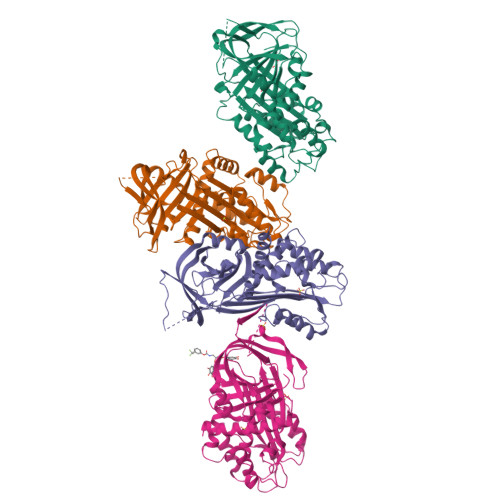
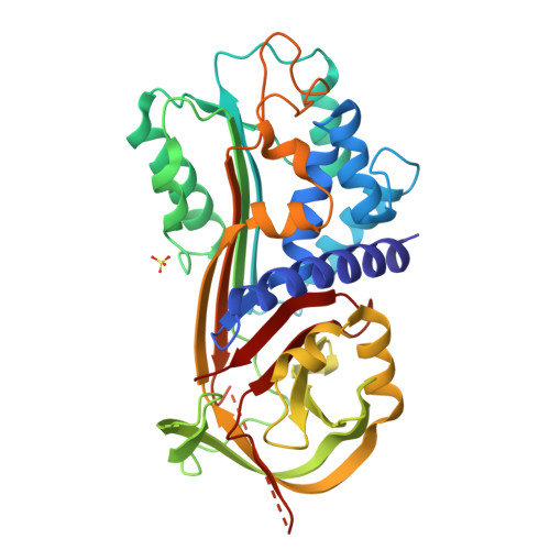
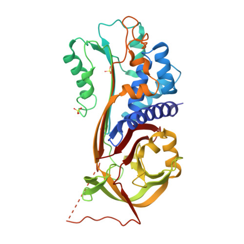
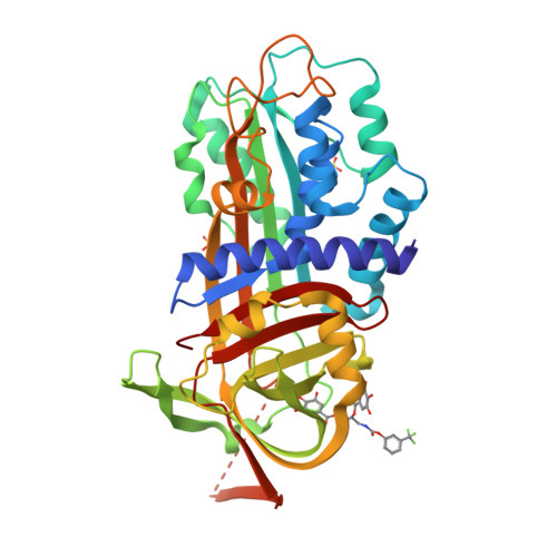
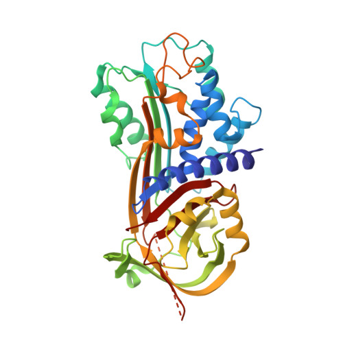
    Plasminogen activator inhibitor type-1 (PAI-1) is a member of the serine protease inhibitor (serpin) family. Excessive PAI-1 activity is associated with human disease, making it an attractive pharmaceutical target. However, like other serpins, PAI-1 has a labile structure, making it a difficult target for the development of small molecule inhibitors, and to date, there are no US Food and Drug Administration-approved small molecule inactivators of any serpins. Here we describe the mechanistic and structural characterization of a high affinity inactivator of PAI-1. This molecule binds to PAI-1 reversibly and acts through an allosteric mechanism that inhibits PAI-1 binding to proteases and to its cofactor vitronectin. The binding site is identified by X-ray crystallography and mutagenesis as a pocket at the interface of β-sheets B and C and α-helix H. A similar pocket is present on other serpins, suggesting that this site could be a common target in this structurally conserved protein family.


  • Organizational Affiliation

    Departments of Pathology and Internal Medicine, Division of Cardiovascular Medicine, University of Michigan Medical School, Ann Arbor, MI 48109.


Macromolecules
Find similar proteins by:  (by identity cutoff)  |  3D Structure
Entity ID: 1
MoleculeChains Sequence LengthOrganismDetailsImage
Plasminogen activator inhibitor 1
A, B, C, D
376Homo sapiensMutation(s): 4 
Gene Names: PAI-1PAI1PLANH1SERPINE1
UniProt & NIH Common Fund Data Resources
Find proteins for P05121 (Homo sapiens)
Explore P05121 
Go to UniProtKB:  P05121
PHAROS:  P05121
GTEx:  ENSG00000106366 
Entity Groups  
Sequence Clusters30% Identity50% Identity70% Identity90% Identity95% Identity100% Identity
UniProt GroupP05121
Sequence Annotations
Expand
  • Reference Sequence
Small Molecules
Ligands 3 Unique
IDChains Name / Formula / InChI Key2D Diagram3D Interactions
96P
Query on 96P

Download Ideal Coordinates CCD File 
L [auth D](2S)-3-({[3-(trifluoromethyl)phenoxy]carbonyl}amino)propane-1,2-diyl bis(3,4,5-trihydroxybenzoate)
C25 H20 F3 N O12
OXWKLJPAKUDPJY-HNNXBMFYSA-N
SO4
Query on SO4

Download Ideal Coordinates CCD File 
E [auth A]
G [auth B]
H [auth C]
I [auth C]
J [auth D]
SULFATE ION
O4 S
QAOWNCQODCNURD-UHFFFAOYSA-L
EDO
Query on EDO

Download Ideal Coordinates CCD File 
F [auth A]1,2-ETHANEDIOL
C2 H6 O2
LYCAIKOWRPUZTN-UHFFFAOYSA-N
Experimental Data & Validation

Experimental Data

  • Method: X-RAY DIFFRACTION
  • Resolution: 2.71 Å
  • R-Value Free:  0.268 (Depositor), 0.290 (DCC) 
  • R-Value Work:  0.239 (Depositor), 0.250 (DCC) 
  • R-Value Observed: 0.241 (Depositor) 
Space Group: P 1
Unit Cell:
Length ( Å )Angle ( ˚ )
a = 65.335α = 91.32
b = 75.317β = 93.49
c = 105.002γ = 115.65
Software Package:
Software NamePurpose
DENZOdata reduction
SCALEPACKdata scaling
BUSTER-TNTrefinement
PDB_EXTRACTdata extraction
MD2data collection
HKL-2000data reduction
HKL-2000data scaling
BUSTERrefinement

Structure Validation

View Full Validation Report


Ligand Structure Quality Assessment 

Created with Raphaël 2.3.0Worse 01 BetterLigand structure goodness of fit to experimental dataBest fitted 96PClick on this verticalbar to view details

Entry History 

Deposition Data

Revision History  (Full details and data files)

  • Version 1.0: 2013-12-25
    Type: Initial release
  • Version 1.1: 2014-01-01
    Changes: Database references
  • Version 1.2: 2017-11-15
    Changes: Advisory, Refinement description
  • Version 1.3: 2024-02-28
    Changes: Advisory, Data collection, Database references, Derived calculations