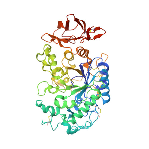
4GQR

Human Pancreatic alpha-amylase in complex with myricetin


Experimental Data Snapshot

  • Method: X-RAY DIFFRACTION
  • Resolution: 1.20 Å
  • R-Value Free: 0.198 
  • R-Value Work: 0.180 

wwPDB Validation   3D Report Full Report


Ligand Structure Quality Assessment 


This is version 2.1 of the entry. See complete history


Literature

Order and disorder: differential structural impacts of myricetin and ethyl caffeate on human amylase, an antidiabetic target.

Williams, L.K.Li, C.Withers, S.G.Brayer, G.D.

(2012) J Med Chem 55: 10177-10186

  • DOI: https://doi.org/10.1021/jm301273u
  • Primary Citation of Related Structures:  
    4GQQ, 4GQR

  • PubMed Abstract: 

    The increasing prevalence of diabetes has accelerated the search for new drugs derived from natural sources. To define the functional features of two such families of compounds, the flavonols and the ethyl caffeates, we have determined the high-resolution structures of representative inhibitors in complex with human pancreatic α-amylase. Myricetin binds at the active site and interacts directly with the catalytic residues despite its bulky planar nature. Notably, it reduces the normal conformational flexibility of the adjacent substrate binding cleft. In contrast, bound ethyl caffeate acts by disordering precisely those polypeptide chain segments that make up the active site binding cleft. It also operates from binding sites far removed from the active site, a property not observed in any other class of human α-amylase inhibitor studied to date. Given the current inadequacy of drugs directed at diabetes, the use of optimized flavonols and ethyl caffeates may present an alternative therapeutic route.


  • Organizational Affiliation

    Department of Biochemistry and Molecular Biology, University of British Columbia , Vancouver, British Columbia V6T 1Z3, Canada.


Macromolecules
Find similar proteins by:  (by identity cutoff)  |  3D Structure
Entity ID: 1
MoleculeChains Sequence LengthOrganismDetailsImage
Pancreatic alpha-amylase496Homo sapiensMutation(s): 0 
Gene Names: AMY2A
EC: 3.2.1.1
UniProt & NIH Common Fund Data Resources
Find proteins for P04746 (Homo sapiens)
Explore P04746 
Go to UniProtKB:  P04746
PHAROS:  P04746
GTEx:  ENSG00000243480 
Entity Groups  
Sequence Clusters30% Identity50% Identity70% Identity90% Identity95% Identity100% Identity
UniProt GroupP04746
Glycosylation
Glycosylation Sites: 1Go to GlyGen: P04746-1
Sequence Annotations
Expand
  • Reference Sequence
Small Molecules
Modified Residues  1 Unique
IDChains TypeFormula2D DiagramParent
PCA
Query on PCA
A
L-PEPTIDE LINKINGC5 H7 N O3GLN
Binding Affinity Annotations 
IDSourceBinding Affinity
MYC PDBBind:  4GQR Ki: 1.10e+5 (nM) from 1 assay(s)
BindingDB:  4GQR IC50: 3.02e+4 (nM) from 1 assay(s)
Experimental Data & Validation

Experimental Data

  • Method: X-RAY DIFFRACTION
  • Resolution: 1.20 Å
  • R-Value Free: 0.198 
  • R-Value Work: 0.180 
  • Space Group: P 21 21 21
Unit Cell:
Length ( Å )Angle ( ˚ )
a = 52.129α = 90
b = 67.373β = 90
c = 129.929γ = 90
Software Package:
Software NamePurpose
Blu-Icedata collection
CNSrefinement
MOSFLMdata reduction
SCALAdata scaling
CNSphasing

Structure Validation

View Full Validation Report


Ligand Structure Quality Assessment 


Entry History 

Deposition Data

Revision History  (Full details and data files)

  • Version 1.0: 2012-10-24
    Type: Initial release
  • Version 1.1: 2012-12-12
    Changes: Database references
  • Version 2.0: 2020-07-29
    Type: Remediation
    Reason: Carbohydrate remediation
    Changes: Data collection, Derived calculations, Polymer sequence, Structure summary
  • Version 2.1: 2024-11-27
    Changes: Data collection, Database references, Structure summary