4KSY | pdb_00004ksy

Crystal structure of STING in complex with cGAMP


Experimental Data Snapshot

  • Method: X-RAY DIFFRACTION
  • Resolution: 1.88 Å
  • R-Value Free: 
    0.181 (Depositor), 0.180 (DCC) 
  • R-Value Work: 
    0.161 (Depositor), 0.160 (DCC) 
  • R-Value Observed: 
    0.163 (Depositor) 

wwPDB Validation   3D Report Full Report


Ligand Structure Quality Assessment 

Created with Raphaël 2.3.0Worse 01 BetterLigand structure goodness of fit to experimental dataBest fitted 1SYClick on this verticalbar to view details

This is version 1.5 of the entry. See complete history


Literature

Cyclic GMP-AMP Containing Mixed Phosphodiester Linkages Is An Endogenous High-Affinity Ligand for STING.

Zhang, X.Shi, H.Wu, J.Zhang, X.Sun, L.Chen, C.Chen, Z.J.

(2013) Mol Cell 51: 226-235

  • DOI: https://doi.org/10.1016/j.molcel.2013.05.022
  • Primary Citation of Related Structures:  
    4KSY

  • PubMed Abstract: 

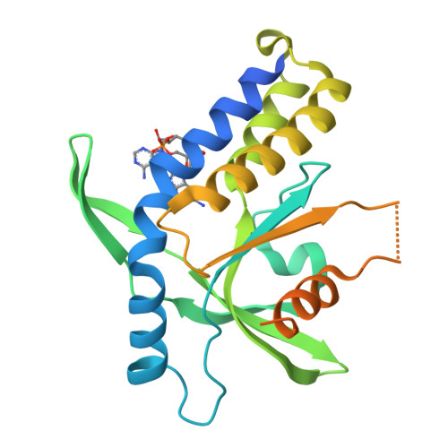
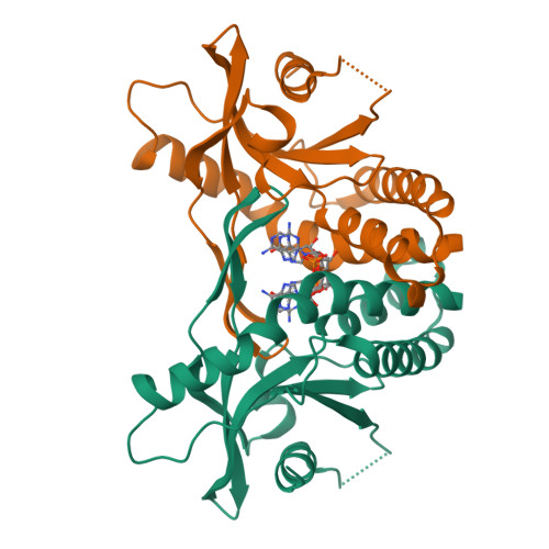
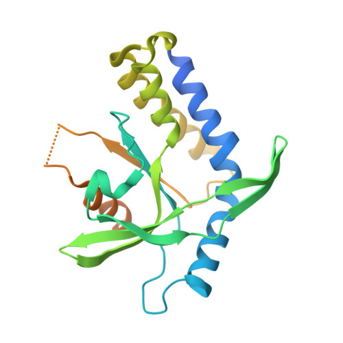
    The presence of microbial or self DNA in the cytoplasm of mammalian cells is a danger signal detected by the DNA sensor cyclic-GMP-AMP (cGAMP) synthase (cGAS), which catalyzes the production of cGAMP that in turn serves as a second messenger to activate innate immune responses. Here we show that endogenous cGAMP in mammalian cells contains two distinct phosphodiester linkages, one between 2'-OH of GMP and 5'-phosphate of AMP, and the other between 3'-OH of AMP and 5'-phosphate of GMP. This molecule, termed 2'3'-cGAMP, is unique in that it binds to the adaptor protein STING with a much greater affinity than cGAMP molecules containing other combinations of phosphodiester linkages. The crystal structure of STING bound to 2'3'-cGAMP revealed the structural basis of this high-affinity binding and a ligand-induced conformational change in STING that may underlie its activation.


  • Organizational Affiliation

    Department of Molecular Biology, University of Texas Southwestern Medical Center, Dallas, TX 75390-9148, USA.


Macromolecules
Find similar proteins by:  (by identity cutoff)  |  3D Structure
Entity ID: 1
MoleculeChains Sequence LengthOrganismDetailsImage
Stimulator of interferon genes protein242Homo sapiensMutation(s): 1 
Gene Names: TMEM173ERISMITASTING
UniProt & NIH Common Fund Data Resources
Find proteins for Q86WV6 (Homo sapiens)
Explore Q86WV6 
Go to UniProtKB:  Q86WV6
PHAROS:  Q86WV6
GTEx:  ENSG00000184584 
Entity Groups  
Sequence Clusters30% Identity50% Identity70% Identity90% Identity95% Identity100% Identity
UniProt GroupQ86WV6
Sequence Annotations
Expand
  • Reference Sequence
Small Molecules
Ligands 1 Unique
IDChains Name / Formula / InChI Key2D Diagram3D Interactions
1SY
Query on 1SY

Download Ideal Coordinates CCD File 
B [auth A]cGAMP
C20 H24 N10 O13 P2
XRILCFTWUCUKJR-INFSMZHSSA-N
Experimental Data & Validation

Experimental Data

  • Method: X-RAY DIFFRACTION
  • Resolution: 1.88 Å
  • R-Value Free:  0.181 (Depositor), 0.180 (DCC) 
  • R-Value Work:  0.161 (Depositor), 0.160 (DCC) 
  • R-Value Observed: 0.163 (Depositor) 
Space Group: C 1 2 1
Unit Cell:
Length ( Å )Angle ( ˚ )
a = 89.522α = 90
b = 77.927β = 96.97
c = 35.974γ = 90
Software Package:
Software NamePurpose
HKL-3000data collection
PHENIXrefinement
HKL-2000data reduction
HKL-2000data scaling

Structure Validation

View Full Validation Report


Ligand Structure Quality Assessment 

Created with Raphaël 2.3.0Worse 01 BetterLigand structure goodness of fit to experimental dataBest fitted 1SYClick on this verticalbar to view details

Entry History 

Deposition Data

Revision History  (Full details and data files)

  • Version 1.0: 2013-06-12
    Type: Initial release
  • Version 1.1: 2013-07-17
    Changes: Database references
  • Version 1.2: 2013-08-07
    Changes: Database references
  • Version 1.3: 2015-08-05
    Changes: Non-polymer description
  • Version 1.4: 2018-06-27
    Changes: Data collection, Database references
  • Version 1.5: 2024-02-28
    Changes: Data collection, Database references, Derived calculations, Structure summary