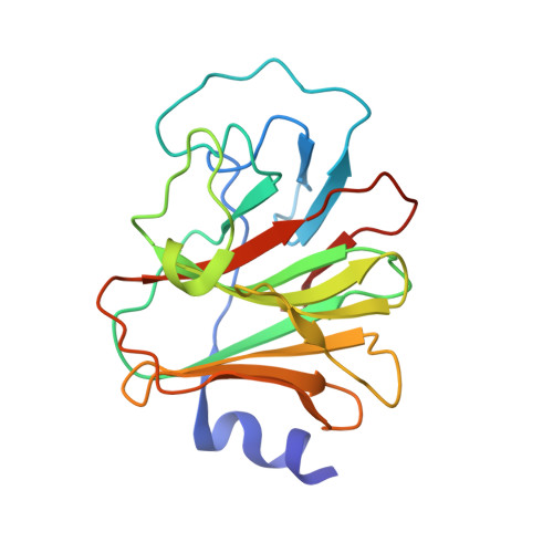
4N7U

Crystal Structure of Intracellular B30.2 Domain of BTN3A1 in Complex with CHDMAPP


Experimental Data Snapshot

  • Method: X-RAY DIFFRACTION
  • Resolution: 1.46 Å
  • R-Value Free: 0.190 
  • R-Value Work: 0.153 
  • R-Value Observed: 0.155 

Starting Model: experimental
View more details

wwPDB Validation   3D Report Full Report


Ligand Structure Quality Assessment 


This is version 1.3 of the entry. See complete history


Literature

The Intracellular B30.2 Domain of Butyrophilin 3A1 Binds Phosphoantigens to Mediate Activation of Human V gamma 9V delta 2 T Cells.

Sandstrom, A.Peigne, C.M.Leger, A.Crooks, J.E.Konczak, F.Gesnel, M.C.Breathnach, R.Bonneville, M.Scotet, E.Adams, E.J.

(2014) Immunity 40: 490-500

  • DOI: https://doi.org/10.1016/j.immuni.2014.03.003
  • Primary Citation of Related Structures:  
    4N7I, 4N7U

  • PubMed Abstract: 

    In humans, Vγ9Vδ2 T cells detect tumor cells and microbial infections, including Mycobacterium tuberculosis, through recognition of small pyrophosphate containing organic molecules known as phosphoantigens (pAgs). Key to pAg-mediated activation of Vγ9Vδ2 T cells is the butyrophilin 3A1 (BTN3A1) protein that contains an intracellular B30.2 domain critical to pAg reactivity. Here, we have demonstrated through structural, biophysical, and functional approaches that the intracellular B30.2 domain of BTN3A1 directly binds pAg through a positively charged surface pocket. Charge reversal of pocket residues abrogates binding and Vγ9Vδ2 T cell activation. We have also identified a gain-of-function mutation within this pocket that, when introduced into the B30.2 domain of the nonstimulatory BTN3A3 isoform, transfers pAg binding ability and Vγ9Vδ2 T cell activation. These studies demonstrate that internal sensing of changes in pAg metabolite concentrations by BTN3A1 molecules is a critical step in Vγ9Vδ2 T cell detection of infection and tumorigenesis.


  • Organizational Affiliation

    Department of Biochemistry and Molecular Biology, University of Chicago, Chicago, IL 60637, USA.


Macromolecules
Find similar proteins by:  (by identity cutoff)  |  3D Structure
Entity ID: 1
MoleculeChains Sequence LengthOrganismDetailsImage
Butyrophilin subfamily 3 member A1190Homo sapiensMutation(s): 0 
Gene Names: BTF5BTN3A1
UniProt & NIH Common Fund Data Resources
Find proteins for O00481 (Homo sapiens)
Explore O00481 
Go to UniProtKB:  O00481
PHAROS:  O00481
GTEx:  ENSG00000026950 
Entity Groups  
Sequence Clusters30% Identity50% Identity70% Identity90% Identity95% Identity100% Identity
UniProt GroupO00481
Sequence Annotations
Expand
  • Reference Sequence
Small Molecules
Ligands 2 Unique
IDChains Name / Formula / InChI Key2D Diagram3D Interactions
2JA
Query on 2JA

Download Ideal Coordinates CCD File 
B [auth A][(E)-4-methyl-5-oxidanyl-pent-3-enyl]-phosphonooxy-phosphinic acid
C6 H14 O7 P2
YYYUHBYKZCBOPY-ZZXKWVIFSA-N
GOL
Query on GOL

Download Ideal Coordinates CCD File 
C [auth A]GLYCEROL
C3 H8 O3
PEDCQBHIVMGVHV-UHFFFAOYSA-N
Binding Affinity Annotations 
IDSourceBinding Affinity
2JA BindingDB:  4N7U IC50: 7870 (nM) from 1 assay(s)
EC50: min: 2, max: 86 (nM) from 2 assay(s)
Experimental Data & Validation

Experimental Data

  • Method: X-RAY DIFFRACTION
  • Resolution: 1.46 Å
  • R-Value Free: 0.190 
  • R-Value Work: 0.153 
  • R-Value Observed: 0.155 
  • Space Group: P 2 21 21
Unit Cell:
Length ( Å )Angle ( ˚ )
a = 38.98α = 90
b = 44.952β = 90
c = 125.262γ = 90
Software Package:
Software NamePurpose
SCALEPACKdata scaling
PHENIXrefinement
PDB_EXTRACTdata extraction
XDSdata scaling
XSCALEdata scaling
PHASERphasing

Structure Validation

View Full Validation Report


Ligand Structure Quality Assessment 


Entry History 

Deposition Data

Revision History  (Full details and data files)

  • Version 1.0: 2014-04-23
    Type: Initial release
  • Version 1.1: 2014-05-14
    Changes: Database references
  • Version 1.2: 2023-09-20
    Changes: Data collection, Database references, Derived calculations, Refinement description
  • Version 1.3: 2024-03-13
    Changes: Source and taxonomy, Structure summary