4NWX | pdb_00004nwx

Crystal structure of phosphoglycerate mutase from Staphylococcus aureus in 2-phosphoglyceric acid bound form


Experimental Data Snapshot

  • Method: X-RAY DIFFRACTION
  • Resolution: 2.01 Å
  • R-Value Free: 
    0.216 (Depositor), 0.220 (DCC) 
  • R-Value Work: 
    0.168 (Depositor), 0.180 (DCC) 
  • R-Value Observed: 
    0.170 (Depositor) 

Starting Model: experimental
View more details

wwPDB Validation   3D Report Full Report


Ligand Structure Quality Assessment 

Created with Raphaël 2.3.0Worse 01 BetterLigand structure goodness of fit to experimental dataBest fitted 2PGClick on this verticalbar to view details

This is version 1.3 of the entry. See complete history


Literature

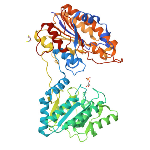
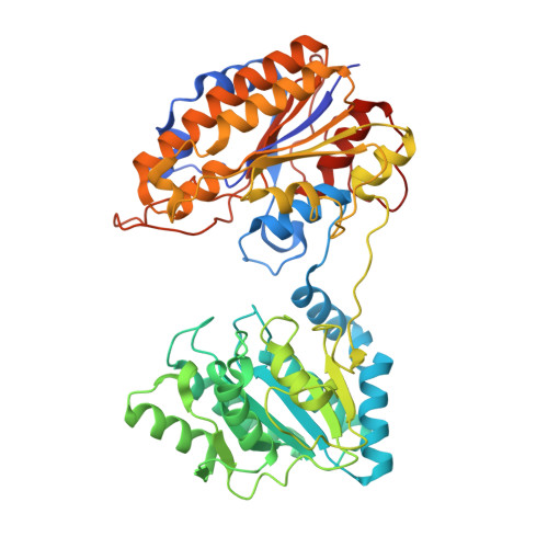
Complete catalytic cycle of cofactor-independent phosphoglycerate mutase involves a spring-loaded mechanism

Roychowdhury, A.Kundu, A.Bose, M.Gujar, A.Mukherjee, S.Das, A.K.

(2015) FEBS J 282: 1097-1110

  • DOI: https://doi.org/10.1111/febs.13205
  • Primary Citation of Related Structures:  
    4MY4, 4NWJ, 4NWX

  • PubMed Abstract: 

    Cofactor-independent phosphoglycerate mutase (iPGM), an important enzyme in glycolysis and gluconeogenesis, catalyses the isomerization of 2- and 3-phosphoglycerates by an Mn(2+)-dependent phospho-transfer mechanism via a phospho-enzyme intermediate. Crystal structures of bi-domain iPGM from Staphylococcus aureus, together with substrate-bound forms, have revealed a new conformation of the enzyme, representing an intermediate state of domain movement. The substrate-binding site and the catalytic site are present in two distinct domains in the intermediate form. X-ray crystallography complemented by simulated dynamics has enabled delineation of the complete catalytic cycle, which includes binding of the substrate, followed by its positioning into the catalytic site, phospho-transfer and finally product release. The present work describes a novel mechanism of domain movement controlled by a hydrophobic patch that is exposed on domain closure and acts like a spring to keep the protein in open conformation. Domain closing occurs after substrate binding, and is essential for phospho-transfer, whereas the open conformation is a prerequisite for efficient substrate binding and product dissociation. A new model of catalysis has been proposed by correlating the hinge-bending motion with the phospho-transfer mechanism.


  • Organizational Affiliation

    Department of Biotechnology, Indian Institute of Technology, Kharagpur, India.


Macromolecules
Find similar proteins by:  (by identity cutoff)  |  3D Structure
Entity ID: 1
MoleculeChains Sequence LengthOrganismDetailsImage
2,3-bisphosphoglycerate-independent phosphoglycerate mutase513Staphylococcus aureus subsp. aureus NCTC 8325Mutation(s): 0 
Gene Names: gpmISAOUHSC_00798
EC: 5.4.2.12
UniProt
Find proteins for Q2G029 (Staphylococcus aureus (strain NCTC 8325 / PS 47))
Explore Q2G029 
Go to UniProtKB:  Q2G029
Entity Groups  
Sequence Clusters30% Identity50% Identity70% Identity90% Identity95% Identity100% Identity
UniProt GroupQ2G029
Sequence Annotations
Expand
  • Reference Sequence
Experimental Data & Validation

Experimental Data

  • Method: X-RAY DIFFRACTION
  • Resolution: 2.01 Å
  • R-Value Free:  0.216 (Depositor), 0.220 (DCC) 
  • R-Value Work:  0.168 (Depositor), 0.180 (DCC) 
  • R-Value Observed: 0.170 (Depositor) 
Space Group: P 21 21 21
Unit Cell:
Length ( Å )Angle ( ˚ )
a = 73.639α = 90
b = 83.206β = 90
c = 89.073γ = 90
Software Package:
Software NamePurpose
SCALAdata scaling
MOLREPphasing
REFMACrefinement
PDB_EXTRACTdata extraction
Structuredata collection
XDSdata reduction

Structure Validation

View Full Validation Report


Ligand Structure Quality Assessment 

Created with Raphaël 2.3.0Worse 01 BetterLigand structure goodness of fit to experimental dataBest fitted 2PGClick on this verticalbar to view details

Entry History 

Deposition Data

Revision History  (Full details and data files)

  • Version 1.0: 2015-01-14
    Type: Initial release
  • Version 1.1: 2015-02-11
    Changes: Database references
  • Version 1.2: 2015-04-08
    Changes: Database references
  • Version 1.3: 2023-11-08
    Changes: Data collection, Database references, Derived calculations, Refinement description