Biochemical and Structural Analysis of Inhibitors Targeting the ADC-7 Cephalosporinase of Acinetobacter baumannii.
Powers, R.A., Swanson, H.C., Taracila, M.A., Florek, N.W., Romagnoli, C., Caselli, E., Prati, F., Bonomo, R.A., Wallar, B.J.(2014) Biochemistry 53: 7670-7679
- PubMed: 25380506
- DOI: https://doi.org/10.1021/bi500887n
- Primary Citation of Related Structures:
4U0T, 4U0X - PubMed Abstract:
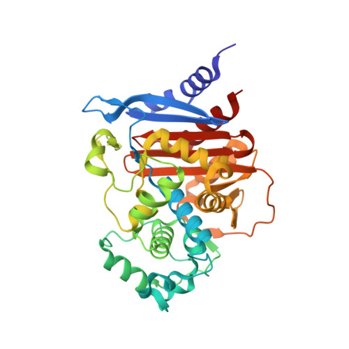
β-Lactam resistance in Acinetobacter baumannii presents one of the greatest challenges to contemporary antimicrobial chemotherapy. Much of this resistance to cephalosporins derives from the expression of the class C β-lactamase enzymes, known as Acinetobacter-derived cephalosporinases (ADCs). Currently, β-lactamase inhibitors are structurally similar to β-lactam substrates and are not effective inactivators of this class C cephalosporinase. Herein, two boronic acid transition state inhibitors (BATSIs S02030 and SM23) that are chemically distinct from β-lactams were designed and tested for inhibition of ADC enzymes. BATSIs SM23 and S02030 bind with high affinity to ADC-7, a chromosomal cephalosporinase from Acinetobacter baumannii (Ki = 21.1 ± 1.9 nM and 44.5 ± 2.2 nM, respectively). The X-ray crystal structures of ADC-7 were determined in both the apo form (1.73 Å resolution) and in complex with S02030 (2.0 Å resolution). In the complex, S02030 makes several canonical interactions: the O1 oxygen of S02030 is bound in the oxyanion hole, and the R1 amide group makes key interactions with conserved residues Asn152 and Gln120. In addition, the carboxylate group of the inhibitor is meant to mimic the C3/C4 carboxylate found in β-lactams. The C3/C4 carboxylate recognition site in class C enzymes is comprised of Asn346 and Arg349 (AmpC numbering), and these residues are conserved in ADC-7. Interestingly, in the ADC-7/S02030 complex, the inhibitor carboxylate group is observed to interact with Arg340, a residue that distinguishes ADC-7 from the related class C enzyme AmpC. A thermodynamic analysis suggests that ΔH driven compounds may be optimized to generate new lead agents. The ADC-7/BATSI complex provides insight into recognition of non-β-lactam inhibitors by ADC enzymes and offers a starting point for the structure-based optimization of this class of novel β-lactamase inhibitors against a key resistance target.
Organizational Affiliation:
Department of Chemistry, Grand Valley State University , 1 Campus Drive, Allendale, Michigan 49401, United States.


