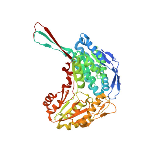
4WP7

Structure of human ALDH1A1 with inhibitor CM026


Experimental Data Snapshot

  • Method: X-RAY DIFFRACTION
  • Resolution: 1.80 Å
  • R-Value Free: 0.224 
  • R-Value Work: 0.185 
  • R-Value Observed: 0.187 

wwPDB Validation   3D Report Full Report


Ligand Structure Quality Assessment 


This is version 1.4 of the entry. See complete history


Literature

Characterization of Two Distinct Structural Classes of Selective Aldehyde Dehydrogenase 1A1 Inhibitors.

Morgan, C.A.Hurley, T.D.

(2015) J Med Chem 58: 1964-1975

  • DOI: https://doi.org/10.1021/jm501900s
  • Primary Citation of Related Structures:  
    4WP7, 4WPN, 4X4L

  • PubMed Abstract: 

    Aldehyde dehydrogenases (ALDH) catalyze the irreversible oxidation of aldehydes to their corresponding carboxylic acid. Alterations in ALDH1A1 activity are associated with such diverse diseases as cancer, Parkinson's disease, obesity, and cataracts. Inhibitors of ALDH1A1 could aid in illuminating the role of this enzyme in disease processes. However, there are no commercially available selective inhibitors for ALDH1A1. Here we characterize two distinct chemical classes of inhibitors that are selective for human ALDH1A1 compared to eight other ALDH isoenzymes. The prototypical members of each structural class, CM026 and CM037, exhibit submicromolar inhibition constants but have different mechanisms of inhibition. The crystal structures of these compounds bound to ALDH1A1 demonstrate that they bind within the aldehyde binding pocket of ALDH1A1 and exploit the presence of a unique glycine residue to achieve their selectivity. These two novel and selective ALDH1A1 inhibitors may serve as chemical tools to better understand the contributions of ALDH1A1 to normal biology and to disease states.


  • Organizational Affiliation

    Department of Biochemistry and Molecular Biology Indiana University School of Medicine 635 Barnhill Drive, Indianapolis, Indiana 46202, United States.


Macromolecules
Find similar proteins by:  (by identity cutoff)  |  3D Structure
Entity ID: 1
MoleculeChains Sequence LengthOrganismDetailsImage
Retinal dehydrogenase 1501Homo sapiensMutation(s): 1 
Gene Names: ALDH1A1ALDCALDH1PUMB1
EC: 1.2.1.36 (PDB Primary Data), 1.2.1.19 (UniProt), 1.2.1.28 (UniProt), 1.2.1.3 (UniProt)
UniProt & NIH Common Fund Data Resources
Find proteins for P00352 (Homo sapiens)
Explore P00352 
Go to UniProtKB:  P00352
PHAROS:  P00352
GTEx:  ENSG00000165092 
Entity Groups  
Sequence Clusters30% Identity50% Identity70% Identity90% Identity95% Identity100% Identity
UniProt GroupP00352
Sequence Annotations
Expand
  • Reference Sequence
Small Molecules
Binding Affinity Annotations 
IDSourceBinding Affinity
3SR BindingDB:  4WP7 Ki: min: 720, max: 800 (nM) from 2 assay(s)
IC50: min: 800, max: 1210 (nM) from 2 assay(s)
Experimental Data & Validation

Experimental Data

  • Method: X-RAY DIFFRACTION
  • Resolution: 1.80 Å
  • R-Value Free: 0.224 
  • R-Value Work: 0.185 
  • R-Value Observed: 0.187 
  • Space Group: P 4 2 2
Unit Cell:
Length ( Å )Angle ( ˚ )
a = 109.054α = 90
b = 109.054β = 90
c = 83.225γ = 90
Software Package:
Software NamePurpose
REFMACrefinement

Structure Validation

View Full Validation Report


Ligand Structure Quality Assessment 


Entry History & Funding Information

Deposition Data


Funding OrganizationLocationGrant Number
National Institutes of Health/National Institute on Alcohol Abuse and Alcoholism (NIH/NIAAA)United StatesR01AA018123

Revision History  (Full details and data files)

  • Version 1.0: 2015-02-11
    Type: Initial release
  • Version 1.1: 2015-03-11
    Changes: Database references
  • Version 1.2: 2017-09-06
    Changes: Author supporting evidence, Database references, Derived calculations, Other, Source and taxonomy, Structure summary
  • Version 1.3: 2019-12-11
    Changes: Author supporting evidence
  • Version 1.4: 2023-12-27
    Changes: Data collection, Database references, Derived calculations, Refinement description