4TX9 | pdb_00004tx9

Crystal structure of HisAp from Streptomyces sviceus with degraded ProFAR


Experimental Data Snapshot

  • Method: X-RAY DIFFRACTION
  • Resolution: 1.60 Å
  • R-Value Free: 
    0.180 (Depositor), 0.190 (DCC) 
  • R-Value Work: 
    0.152 (Depositor), 0.170 (DCC) 
  • R-Value Observed: 
    0.153 (Depositor) 

wwPDB Validation   3D Report Full Report


Ligand Structure Quality Assessment 

Created with Raphaël 2.3.0Worse 01 BetterLigand structure goodness of fit to experimental dataBest fitted AMZClick on this verticalbar to view details

This is version 1.6 of the entry. See complete history


Literature

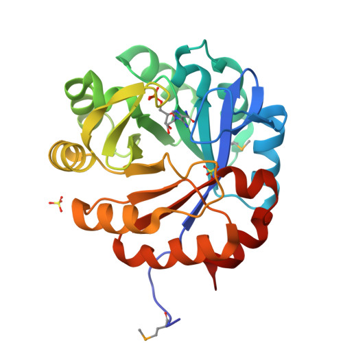
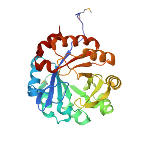
Co-occurrence of analogous enzymes determines evolution of a novel ( beta alpha )8-isomerase sub-family after non-conserved mutations in flexible loop.

Verduzco-Castro, E.A.Michalska, K.Endres, M.Juarez-Vazquez, A.L.Noda-Garcia, L.Chang, C.Henry, C.S.Babnigg, G.Joachimiak, A.Barona-Gomez, F.

(2016) Biochem J 473: 1141-1152

  • DOI: https://doi.org/10.1042/BJ20151271
  • Primary Citation of Related Structures:  
    4TX9, 4U28, 4W9T, 4WUI, 4X9S, 5DN1

  • PubMed Abstract: 

    We investigate the evolution of co-occurring analogous enzymes involved in L-tryptophan and L-histidine biosynthesis in Actinobacteria Phylogenetic analysis of trpF homologues, a missing gene in certain clades of this lineage whose absence is complemented by a dual-substrate HisA homologue, termed PriA, found that they fall into three categories: (i) trpF-1, an L-tryptophan biosynthetic gene horizontally acquired by certain Corynebacterium species; (ii) trpF-2, a paralogue known to be involved in synthesizing a pyrrolopyrrole moiety and (iii) trpF-3, a variable non-conserved orthologue of trpF-1 We previously investigated the effect of trpF-1 upon the evolution of PriA substrate specificity, but nothing is known about the relationship between trpF-3 and priA After in vitro steady-state enzyme kinetics we found that trpF-3 encodes a phosphoribosyl anthranilate isomerase. However, mutation of this gene in Streptomyces sviceus did not lead to auxothrophy, as expected from the biosynthetic role of trpF-1 Biochemical characterization of a dozen co-occurring TrpF-2 or TrpF-3, with PriA homologues, explained the prototrophic phenotype, and unveiled an enzyme activity trade-off between TrpF and PriA. X-ray structural analysis suggests that the function of these PriA homologues is mediated by non-conserved mutations in the flexible L5 loop, which may be responsible for different substrate affinities. Thus, the PriA homologues that co-occur with TrpF-3 represent a novel enzyme family, termed PriB, which evolved in response to PRA isomerase activity. The characterization of co-occurring enzymes provides insights into the influence of functional redundancy on the evolution of enzyme function, which could be useful for enzyme functional annotation.


  • Organizational Affiliation

    Evolution of Metabolic Diversity Laboratory, Unidad de Genómica Avanzada (Langebio), Cinvestav-IPN, Irapuato, CP36821, México.


Macromolecules
Find similar proteins by:  (by identity cutoff)  |  3D Structure
Entity ID: 1
MoleculeChains Sequence LengthOrganismDetailsImage
Phosphoribosyl isomerase A248Streptomyces sviceus ATCC 29083Mutation(s): 0 
Gene Names: priAhisASSEG_10012
EC: 5.3.1.16 (UniProt), 5.3.1.24 (UniProt)
UniProt
Find proteins for B5I4P8 (Streptomyces sviceus (strain ATCC 29083 / DSM 924 / JCM 4929 / NBRC 13980 / NCIMB 11184 / NRRL 5439 / UC 5370))
Explore B5I4P8 
Go to UniProtKB:  B5I4P8
Entity Groups  
Sequence Clusters30% Identity50% Identity70% Identity90% Identity95% Identity100% Identity
UniProt GroupB5I4P8
Sequence Annotations
Expand
  • Reference Sequence
Small Molecules
Experimental Data & Validation

Experimental Data

  • Method: X-RAY DIFFRACTION
  • Resolution: 1.60 Å
  • R-Value Free:  0.180 (Depositor), 0.190 (DCC) 
  • R-Value Work:  0.152 (Depositor), 0.170 (DCC) 
  • R-Value Observed: 0.153 (Depositor) 
Space Group: P 43 21 2
Unit Cell:
Length ( Å )Angle ( ˚ )
a = 72.546α = 90
b = 72.546β = 90
c = 143.286γ = 90
Software Package:
Software NamePurpose
PHENIXrefinement
HKL-3000phasing
HKL-3000data collection
HKL-3000data scaling
HKL-3000data reduction
SHELXmodel building
MLPHAREphasing
DMmodel building
ARPmodel building
Cootmodel building

Structure Validation

View Full Validation Report


Ligand Structure Quality Assessment 

Created with Raphaël 2.3.0Worse 01 BetterLigand structure goodness of fit to experimental dataBest fitted AMZClick on this verticalbar to view details

Entry History & Funding Information

Deposition Data


Funding OrganizationLocationGrant Number
National Institutes of Health/National Institute of General Medical Sciences (NIH/NIGMS)United StatesGM094585

Revision History  (Full details and data files)

  • Version 1.0: 2014-08-06
    Type: Initial release
  • Version 1.1: 2015-02-04
    Changes: Derived calculations
  • Version 1.2: 2017-09-20
    Changes: Author supporting evidence, Database references, Derived calculations, Other, Refinement description, Source and taxonomy
  • Version 1.3: 2017-11-08
    Changes: Database references
  • Version 1.4: 2019-12-25
    Changes: Author supporting evidence
  • Version 1.5: 2023-12-27
    Changes: Data collection, Database references, Refinement description
  • Version 1.6: 2024-11-06
    Changes: Structure summary