5X3D | pdb_00005x3d

Crystal structure of HEP-CMP-bound form of cytidylyltransferase (CyTase) domain of Fom1 from Streptomyces wedmorensis


Experimental Data Snapshot

  • Method: X-RAY DIFFRACTION
  • Resolution: 1.93 Å
  • R-Value Free: 
    0.234 (Depositor), 0.240 (DCC) 
  • R-Value Work: 
    0.191 (Depositor), 0.200 (DCC) 
  • R-Value Observed: 
    0.193 (Depositor) 

wwPDB Validation   3D Report Full Report


Ligand Structure Quality Assessment 

Created with Raphaël 2.3.0Worse 01 BetterLigand structure goodness of fit to experimental dataBest fitted 7XLClick on this verticalbar to view details

This is version 1.1 of the entry. See complete history


Literature

Fosfomycin Biosynthesis via Transient Cytidylylation of 2-Hydroxyethylphosphonate by the Bifunctional Fom1 Enzyme

Cho, S.H.Kim, S.Y.Tomita, T.Shiraishi, T.Park, J.S.Sato, S.Kudo, F.Eguchi, T.Funa, N.Nishiyama, M.Kuzuyama, T.

(2017) ACS Chem Biol 12: 2209-2215

  • DOI: https://doi.org/10.1021/acschembio.7b00419
  • Primary Citation of Related Structures:  
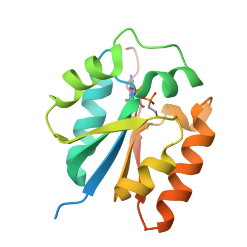
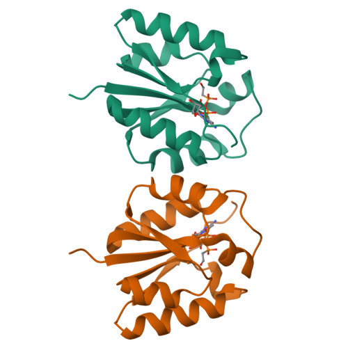
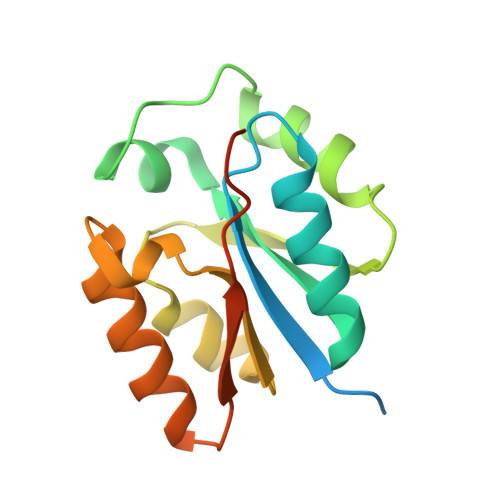
    5X3D

  • PubMed Abstract: 

    Fosfomycin is a wide-spectrum phosphonate antibiotic that is used clinically to treat cystitis, tympanitis, etc. Its biosynthesis starts with the formation of a carbon-phosphorus bond catalyzed by the phosphoenolpyruvate phosphomutase Fom1. We identified an additional cytidylyltransferase (CyTase) domain at the Fom1 N-terminus in addition to the phosphoenolpyruvate phosphomutase domain at the Fom1 C-terminus. Here, we demonstrate that Fom1 is bifunctional and that the Fom1 CyTase domain catalyzes the cytidylylation of the 2-hydroxyethylphosphonate (HEP) intermediate to produce cytidylyl-HEP. On the basis of this new function of Fom1, we propose a revised fosfomycin biosynthetic pathway that involves the transient CMP-conjugated intermediate. The identification of a biosynthetic mechanism via such transient cytidylylation of a biosynthetic intermediate fundamentally advances the understanding of phosphonate biosynthesis in nature. The crystal structure of the cytidylyl-HEP-bound CyTase domain provides a basis for the substrate specificity and reveals unique catalytic elements not found in other members of the CyTase family.


  • Organizational Affiliation

    Biotechnology Research Center, The University of Tokyo , 1-1-1 Yayoi, Bunkyo-ku, Tokyo 113-8657, Japan.


Macromolecules
Find similar proteins by:  (by identity cutoff)  |  3D Structure
Entity ID: 1
MoleculeChains Sequence LengthOrganismDetailsImage
Phosphoenolpyruvate phosphomutase160Streptomyces wedmorensisMutation(s): 0 
Gene Names: Fom1(N)
EC: 5.4.2.9 (UniProt), 2.7.7.104 (UniProt)
UniProt
Find proteins for P96074 (Streptomyces wedmorensis)
Explore P96074 
Go to UniProtKB:  P96074
Entity Groups  
Sequence Clusters30% Identity50% Identity70% Identity90% Identity95% Identity100% Identity
UniProt GroupP96074
Sequence Annotations
Expand
  • Reference Sequence
Small Molecules
Ligands 1 Unique
IDChains Name / Formula / InChI Key2D Diagram3D Interactions
7XL
Query on 7XL

Download Ideal Coordinates CCD File 
B [auth A][[(2R,3S,4R,5R)-5-(4-azanyl-2-oxidanylidene-pyrimidin-1-yl)-3,4-bis(oxidanyl)oxolan-2-yl]methoxy-oxidanyl-phosphoryl]oxy-(2-hydroxyethyl)phosphinic acid
C11 H19 N3 O11 P2
ODFOOQGQRDVSPW-PEBGCTIMSA-N
Experimental Data & Validation

Experimental Data

  • Method: X-RAY DIFFRACTION
  • Resolution: 1.93 Å
  • R-Value Free:  0.234 (Depositor), 0.240 (DCC) 
  • R-Value Work:  0.191 (Depositor), 0.200 (DCC) 
  • R-Value Observed: 0.193 (Depositor) 
Space Group: P 65 2 2
Unit Cell:
Length ( Å )Angle ( ˚ )
a = 71.909α = 90
b = 71.909β = 90
c = 109.507γ = 120
Software Package:
Software NamePurpose
REFMACrefinement
HKL-2000data reduction
HKL-2000data scaling
SOLVEphasing

Structure Validation

View Full Validation Report


Ligand Structure Quality Assessment 

Created with Raphaël 2.3.0Worse 01 BetterLigand structure goodness of fit to experimental dataBest fitted 7XLClick on this verticalbar to view details

Entry History 

Deposition Data

Revision History  (Full details and data files)

  • Version 1.0: 2017-09-13
    Type: Initial release
  • Version 1.1: 2024-03-27
    Changes: Data collection, Database references