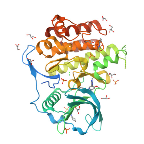
Crystal structure of the FLT3 kinase bound to a small molecule inhibitor
Thomas, C.J.To be published.
Experimental Data Snapshot
Entity ID: 1 | |||||
|---|---|---|---|---|---|
| Molecule | Chains | Sequence Length | Organism | Details | Image |
| Receptor-type tyrosine-protein kinase FLT3 | 370 | Homo sapiens | Mutation(s): 0 Gene Names: FLT3, CD135, FLK2, STK1 EC: 2.7.10.1 | 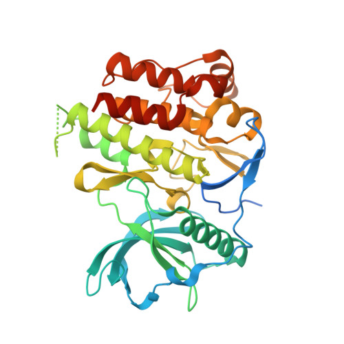 | |
UniProt & NIH Common Fund Data Resources | |||||
Find proteins for P36888 (Homo sapiens) Explore P36888 Go to UniProtKB: P36888 | |||||
PHAROS: P36888 GTEx: ENSG00000122025 | |||||
Entity Groups | |||||
| Sequence Clusters | 30% Identity50% Identity70% Identity90% Identity95% Identity100% Identity | ||||
| UniProt Group | P36888 | ||||
Sequence AnnotationsExpand | |||||
| |||||
| Ligands 5 Unique | |||||
|---|---|---|---|---|---|
| ID | Chains | Name / Formula / InChI Key | 2D Diagram | 3D Interactions | |
| A9R Query on A9R | B [auth A] | 7-methoxy-6-(1-methyl-1H-pyrazol-4-yl)-3-(pyridin-2-yl)imidazo[1,2-a]pyridine C17 H15 N5 O QKGWUZMVBVLWES-UHFFFAOYSA-N |  | ||
| CXS Query on CXS | G [auth A], H [auth A] | 3-CYCLOHEXYL-1-PROPYLSULFONIC ACID C9 H19 N O3 S PJWWRFATQTVXHA-UHFFFAOYSA-N |  | ||
| PO4 Query on PO4 | C [auth A], D [auth A], E [auth A], F [auth A] | PHOSPHATE ION O4 P NBIIXXVUZAFLBC-UHFFFAOYSA-K |  | ||
| GOL Query on GOL | I [auth A] J [auth A] K [auth A] L [auth A] M [auth A] | GLYCEROL C3 H8 O3 PEDCQBHIVMGVHV-UHFFFAOYSA-N |  | ||
| CL Query on CL | S [auth A], T [auth A], U [auth A] | CHLORIDE ION Cl VEXZGXHMUGYJMC-UHFFFAOYSA-M |  | ||
| Length ( Å ) | Angle ( ˚ ) |
|---|---|
| a = 81.641 | α = 90 |
| b = 81.641 | β = 90 |
| c = 147.843 | γ = 90 |
| Software Name | Purpose |
|---|---|
| REFMAC | refinement |
| XDS | data reduction |
| Aimless | data scaling |
| PHASER | phasing |