Segmentation and Comparative Modeling in an 8.6- angstrom Cryo-EM Map of the Singapore Grouper Iridovirus.
Pintilie, G., Chen, D.H., Tran, B.N., Jakana, J., Wu, J., Hew, C.L., Chiu, W.(2019) Structure 27: 1561
- PubMed: 31447288
- DOI: https://doi.org/10.1016/j.str.2019.08.002
- Primary Citation of Related Structures:
6OJN - PubMed Abstract:
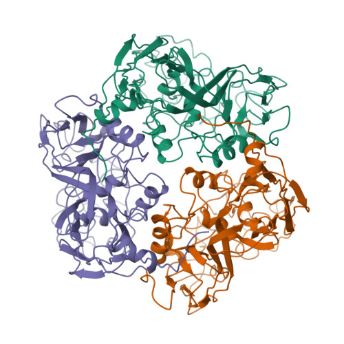
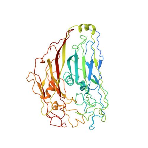
SGIV, or Singapore grouper iridovirus, is a large double-stranded DNA virus, reaching a diameter of 220 nm and packaging a genome of 140 kb. We present a 3D cryoelectron microscopy (cryo-EM) icosahedral reconstruction of SGIV determined at 8.6-Å resolution. It reveals several layers including a T = 247 icosahedral outer coat, anchor proteins, a lipid bilayer, and the encapsidated DNA. A new segmentation tool, iSeg, was applied to extract these layers from the reconstructed map. The outer coat was further segmented into major and minor capsid proteins. None of the proteins extracted by segmentation have known atomic structures. We generated models for the major coat protein using three comparative modeling tools, and evaluated each model using the cryo-EM map. Our analysis reveals a new architecture in the Iridoviridae family of viruses. It shares similarities with others in the same family, e.g., Chilo iridescent virus, but also shows new features of the major and minor capsid proteins.
Organizational Affiliation:
Department of Bioengineering, and of Microbiology and Immunology, Stanford University, Stanford, CA 94305, USA. Electronic address: gregp@slac.stanford.edu.