6S8U | pdb_00006s8u

Structure of the PfEMP1 IT4var13 DBLbeta domain bound to ICAM-1


Experimental Data Snapshot

  • Method: X-RAY DIFFRACTION
  • Resolution: 3.67 Å
  • R-Value Free: 
    0.286 (Depositor), 0.286 (DCC) 
  • R-Value Work: 
    0.240 (Depositor), 0.240 (DCC) 
  • R-Value Observed: 
    0.242 (Depositor) 

wwPDB Validation   3D Report Full Report


Ligand Structure Quality Assessment 

Created with Raphaël 2.3.0Worse 01 BetterLigand structure goodness of fit to experimental dataBest fitted NAGClick on this verticalbar to view details

This is version 2.1 of the entry. See complete history


Literature

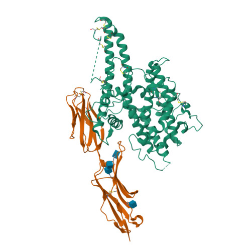
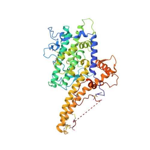
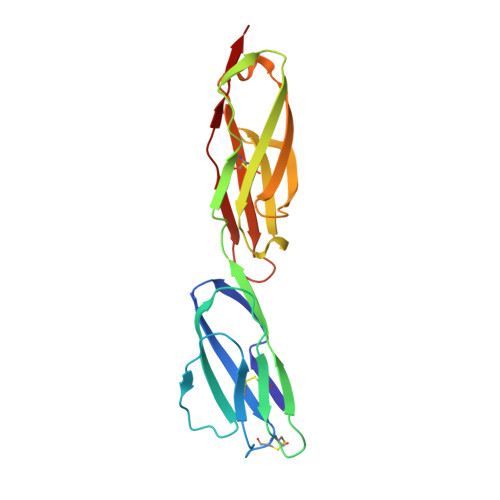
Structural insights into diverse modes of ICAM-1 binding byPlasmodium falciparum-infected erythrocytes.

Lennartz, F.Smith, C.Craig, A.G.Higgins, M.K.

(2019) Proc Natl Acad Sci U S A 116: 20124-20134

  • DOI: https://doi.org/10.1073/pnas.1911900116
  • Primary Citation of Related Structures:  
    6S8T, 6S8U

  • PubMed Abstract: 

    A major determinant of pathogenicity in malaria caused by Plasmodium falciparum is the adhesion of parasite-infected erythrocytes to the vasculature or tissues of infected individuals. This occludes blood flow, leads to inflammation, and increases parasitemia by reducing spleen-mediated clearance of the parasite. This adhesion is mediated by PfEMP1, a multivariant family of around 60 proteins per parasite genome which interact with specific host receptors. One of the most common of these receptors is intracellular adhesion molecule-1 (ICAM-1), which is bound by 2 distinct groups of PfEMP1, A-type and B or C (BC)-type. Here, we present the structure of a domain from a B-type PfEMP1 bound to ICAM-1, revealing a complex binding site. Comparison with the existing structure of an A-type PfEMP1 bound to ICAM-1 shows that the 2 complexes share a globally similar architecture. However, while the A-type PfEMP1 bind ICAM-1 through a highly conserved binding surface, the BC-type PfEMP1 use a binding site that is more diverse in sequence, similar to how PfEMP1 interact with other human receptors. We also show that A- and BC-type PfEMP1 present ICAM-1 at different angles, perhaps influencing the ability of neighboring PfEMP1 domains to bind additional receptors. This illustrates the deep diversity of the PfEMP1 and demonstrates how variations in a single domain architecture can modulate binding to a specific ligand to control function and facilitate immune evasion.


  • Organizational Affiliation

    Department of Biochemistry, University of Oxford, OX1 3QU Oxford, United Kingdom.


Macromolecules
Find similar proteins by:  (by identity cutoff)  |  3D Structure
Entity ID: 1
MoleculeChains Sequence LengthOrganismDetailsImage
Erythrocyte membrane protein 1469Plasmodium falciparumMutation(s): 0 
UniProt
Find proteins for E0A3B3 (Plasmodium falciparum)
Explore E0A3B3 
Go to UniProtKB:  E0A3B3
Entity Groups  
Sequence Clusters30% Identity50% Identity70% Identity90% Identity95% Identity100% Identity
UniProt GroupE0A3B3
Sequence Annotations
Expand
  • Reference Sequence
Find similar proteins by:  (by identity cutoff)  |  3D Structure
Entity ID: 2
MoleculeChains Sequence LengthOrganismDetailsImage
Intercellular adhesion molecule 1186Homo sapiensMutation(s): 0 
Gene Names: ICAM1
UniProt & NIH Common Fund Data Resources
Find proteins for P05362 (Homo sapiens)
Explore P05362 
Go to UniProtKB:  P05362
PHAROS:  P05362
GTEx:  ENSG00000090339 
Entity Groups  
Sequence Clusters30% Identity50% Identity70% Identity90% Identity95% Identity100% Identity
UniProt GroupP05362
Glycosylation
Glycosylation Sites: 3Go to GlyGen: P05362-1
Sequence Annotations
Expand
  • Reference Sequence
Oligosaccharides

Help

Entity ID: 3
MoleculeChains Length2D Diagram Glycosylation3D Interactions
2-acetamido-2-deoxy-beta-D-glucopyranose-(1-4)-2-acetamido-2-deoxy-beta-D-glucopyranose
C
2N-Glycosylation
Glycosylation Resources
GlyTouCan:  G42666HT
GlyCosmos:  G42666HT
GlyGen:  G42666HT
Experimental Data & Validation

Experimental Data

  • Method: X-RAY DIFFRACTION
  • Resolution: 3.67 Å
  • R-Value Free:  0.286 (Depositor), 0.286 (DCC) 
  • R-Value Work:  0.240 (Depositor), 0.240 (DCC) 
  • R-Value Observed: 0.242 (Depositor) 
Space Group: P 65 2 2
Unit Cell:
Length ( Å )Angle ( ˚ )
a = 142.41α = 90
b = 142.41β = 90
c = 224.225γ = 120
Software Package:
Software NamePurpose
PHENIXrefinement
PDB_EXTRACTdata extraction
xia2data reduction
DIALSdata scaling
PHASERphasing

Structure Validation

View Full Validation Report


Ligand Structure Quality Assessment 

Created with Raphaël 2.3.0Worse 01 BetterLigand structure goodness of fit to experimental dataBest fitted NAGClick on this verticalbar to view details

Entry History & Funding Information

Deposition Data


Funding OrganizationLocationGrant Number
Wellcome Trust--

Revision History  (Full details and data files)

  • Version 1.0: 2019-09-25
    Type: Initial release
  • Version 1.1: 2019-10-02
    Changes: Data collection, Database references
  • Version 1.2: 2019-10-09
    Changes: Data collection, Database references
  • Version 2.0: 2020-07-29
    Type: Remediation
    Reason: Carbohydrate remediation
    Changes: Atomic model, Data collection, Derived calculations, Structure summary
  • Version 2.1: 2024-11-13
    Changes: Data collection, Database references, Structure summary