7E7F

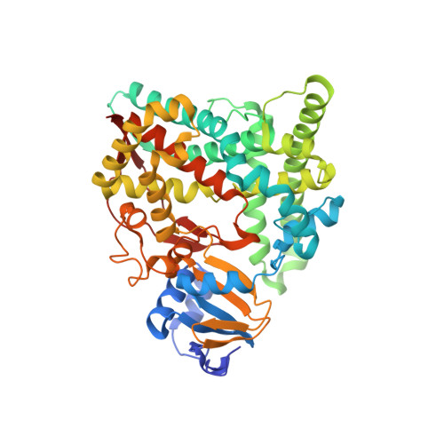
Human CYP11B1 mutant in complex with metyrapone


Experimental Data Snapshot

  • Method: X-RAY DIFFRACTION
  • Resolution: 1.40 Å
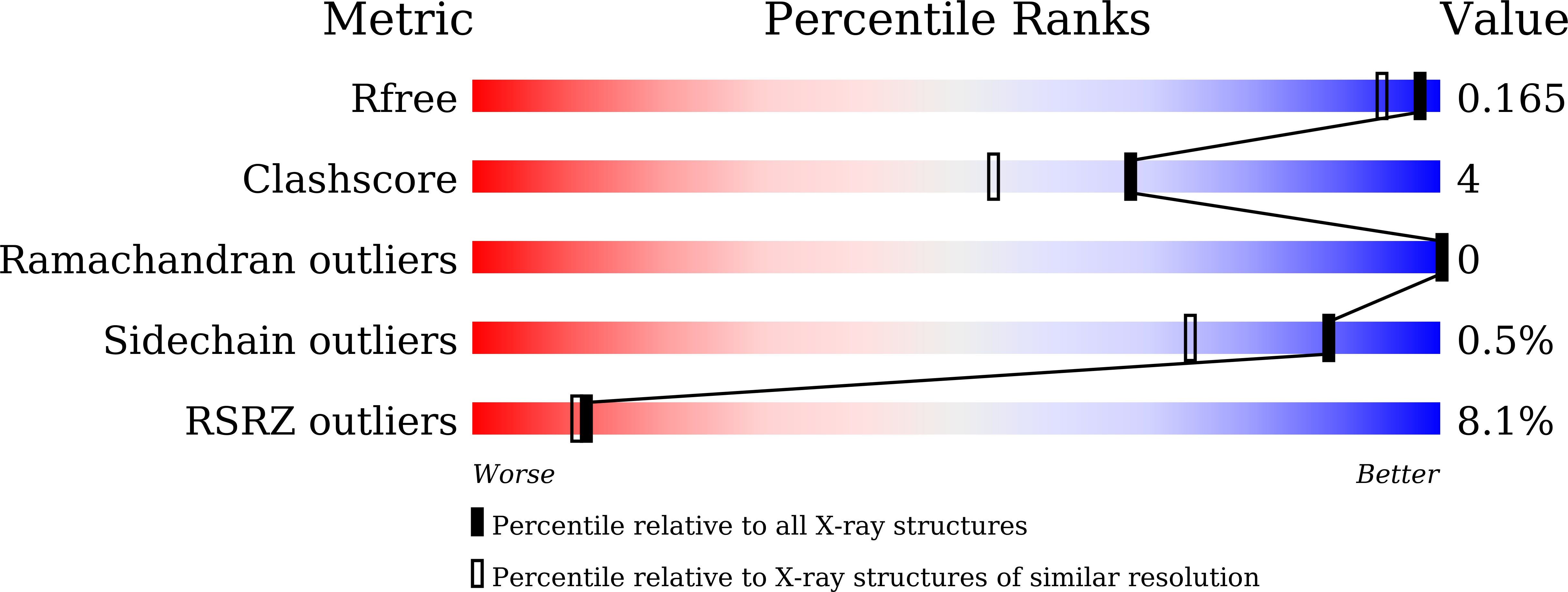
  • R-Value Free: 0.163 
  • R-Value Work: 0.140 
  • R-Value Observed: 0.141 

Starting Model: experimental
View more details

wwPDB Validation   3D Report Full Report


Ligand Structure Quality Assessment 


This is version 1.1 of the entry. See complete history


Literature

Spatially restricted substrate-binding site of cortisol-synthesizing CYP11B1 limits multiple hydroxylations and hinders aldosterone synthesis.

Mukai, K.Sugimoto, H.Kamiya, K.Suzuki, R.Matsuura, T.Hishiki, T.Shimada, H.Shiro, Y.Suematsu, M.Kagawa, N.

(2021) Curr Res Struct Biol 3: 192-205

  • DOI: https://doi.org/10.1016/j.crstbi.2021.08.001
  • Primary Citation of Related Structures:  
    7E7F

  • PubMed Abstract: 

    Human cytochromes P450 11β (CYP11B1) and P450 aldo (CYP11B2) are monooxygenases that synthesize cortisol through steroid 11β-hydroxylation and aldosterone through a three-step process comprising 11β-hydroxylation and two 18-hydroxylations, respectively. CYP11B1 also catalyzes 18-monohydroxylation and 11β,18-dihydroxylation. To study the molecular basis of such catalytic divergence of the two enzymes, we examined a CYP11B1 mutant (Mt-CYP11B1) with amino acid replacements on the distal surface by determining the catalytic activities and crystal structure in the metyrapone-bound form at 1.4-Å resolution. Mt-CY11B1 retained both 11β-hydroxylase and 18-hydroxylase activities of the wild type (Wt-CYP11B1) but lacked 11β,18-dihydroxylase activity. Comparisons of the crystal structure of Mt-CYP11B1 to those of Wt-CYP11B1 and CYP11B2 that were already reported show that the mutation reduced the innermost space putatively surrounding the C3 side of substrate 11-deoxycorticosterone (DOC) bound to Wt-CYP11B1, while the corresponding space in CYP11B2 is enlarged markedly and accessible to bulk water through a channel. Molecular dynamics simulations of their DOC-bound forms supported the above findings and revealed that the enlarged space of CYP11B2 had a hydrogen bonding network involving water molecules that position DOC. Thus, upon positioning 11β-hydroxysteroid for 18-hydroxylation in their substrate-binding sites, steric hindrance could occur more strongly in Mt-CYP11B1 than in Wt-CYP11B1 but less in CYP11B2. Our investigation employing Mt-CYP11B1 sheds light on the divergence in structure and function between CYP11B1 and CYP11B2 and suggests that CYP11B1 with spatially-restricted substrate-binding site serves as 11β-hydroxylase, while CYP11B2 with spatially-extended substrate-binding site successively processes additional 18-hydroxylations to produce aldosterone.


  • Organizational Affiliation

    Department of Biochemistry, Keio University School of Medicine, Shinjuku, Tokyo 160-8582, Japan.


Macromolecules
Find similar proteins by:  (by identity cutoff)  |  3D Structure
Entity ID: 1
MoleculeChains Sequence LengthOrganismDetailsImage
Cytochrome P450 11B1, mitochondrial486Homo sapiensMutation(s): 6 
Gene Names: CYP11B1S11BH
EC: 1.14.15.4
UniProt & NIH Common Fund Data Resources
Find proteins for P15538 (Homo sapiens)
Explore P15538 
Go to UniProtKB:  P15538
PHAROS:  P15538
GTEx:  ENSG00000160882 
Entity Groups  
Sequence Clusters30% Identity50% Identity70% Identity90% Identity95% Identity100% Identity
UniProt GroupP15538
Sequence Annotations
Expand
  • Reference Sequence
Small Molecules
Ligands 4 Unique
IDChains Name / Formula / InChI Key2D Diagram3D Interactions
HEM
Query on HEM

Download Ideal Coordinates CCD File 
B [auth A]PROTOPORPHYRIN IX CONTAINING FE
C34 H32 Fe N4 O4
KABFMIBPWCXCRK-RGGAHWMASA-L
CHD
Query on CHD

Download Ideal Coordinates CCD File 
D [auth A],
E [auth A]
CHOLIC ACID
C24 H40 O5
BHQCQFFYRZLCQQ-OELDTZBJSA-N
MYT (Subject of Investigation/LOI)
Query on MYT

Download Ideal Coordinates CCD File 
C [auth A]METYRAPONE
C14 H14 N2 O
FJLBFSROUSIWMA-UHFFFAOYSA-N
GOL
Query on GOL

Download Ideal Coordinates CCD File 
F [auth A],
G [auth A]
GLYCEROL
C3 H8 O3
PEDCQBHIVMGVHV-UHFFFAOYSA-N
Binding Affinity Annotations 
IDSourceBinding Affinity
MYT BindingDB:  7E7F IC50: min: 5, max: 208 (nM) from 7 assay(s)
Experimental Data & Validation

Experimental Data

  • Method: X-RAY DIFFRACTION
  • Resolution: 1.40 Å
  • R-Value Free: 0.163 
  • R-Value Work: 0.140 
  • R-Value Observed: 0.141 
  • Space Group: P 21 21 21
Unit Cell:
Length ( Å )Angle ( ˚ )
a = 75.18α = 90
b = 84.6β = 90
c = 85.61γ = 90
Software Package:
Software NamePurpose
XSCALEdata scaling
REFMACrefinement
PDB_EXTRACTdata extraction
ARP/wARPdata reduction
PHASERphasing

Structure Validation

View Full Validation Report


Ligand Structure Quality Assessment 


Entry History & Funding Information

Deposition Data


Funding OrganizationLocationGrant Number
Japan Society for the Promotion of Science (JSPS)Japan26461387
Japan Society for the Promotion of Science (JSPS)Japan17K09890

Revision History  (Full details and data files)

  • Version 1.0: 2022-01-05
    Type: Initial release
  • Version 1.1: 2023-11-29
    Changes: Data collection, Refinement description