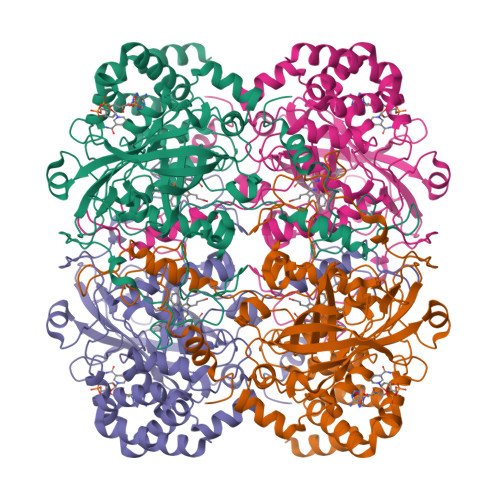
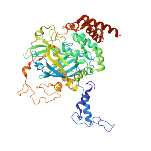
8PVD

Structure of catalase determined by cryoEM at 100 keV


Experimental Data Snapshot

  • Method: ELECTRON MICROSCOPY
  • Resolution: 3.40 Å
  • Aggregation State: PARTICLE 
  • Reconstruction Method: SINGLE PARTICLE 

Starting Model: experimental
View more details

wwPDB Validation   3D Report Full Report


This is version 1.1 of the entry. See complete history


Literature

Structure determination by cryoEM at 100 keV.

McMullan, G.Naydenova, K.Mihaylov, D.Yamashita, K.Peet, M.J.Wilson, H.Dickerson, J.L.Chen, S.Cannone, G.Lee, Y.Hutchings, K.A.Gittins, O.Sobhy, M.A.Wells, T.El-Gomati, M.M.Dalby, J.Meffert, M.Schulze-Briese, C.Henderson, R.Russo, C.J.

(2023) Proc Natl Acad Sci U S A 120: e2312905120-e2312905120

  • DOI: https://doi.org/10.1073/pnas.2312905120
  • Primary Citation of Related Structures:  
    8PV9, 8PVA, 8PVB, 8PVC, 8PVD, 8PVE, 8PVF, 8PVG, 8PVH, 8PVI, 8PVJ

  • PubMed Abstract: 

    Electron cryomicroscopy can, in principle, determine the structures of most biological molecules but is currently limited by access, specimen preparation difficulties, and cost. We describe a purpose-built instrument operating at 100 keV-including advances in electron optics, detection, and processing-that makes structure determination fast and simple at a fraction of current costs. The instrument attains its theoretical performance limits, allowing atomic resolution imaging of gold test specimens and biological molecular structure determination in hours. We demonstrate its capabilities by determining the structures of eleven different specimens, ranging in size from 140 kDa to 2 MDa, using a fraction of the data normally required. CryoEM with a microscope designed specifically for high-efficiency, on-the-spot imaging of biological molecules will expand structural biology to a wide range of previously intractable problems.


  • Organizational Affiliation

    Medical Research Council (MRC) Laboratory of Molecular Biology, Cambridge CB2 0QH, United Kingdom.


Macromolecules
Find similar proteins by:  (by identity cutoff)  |  3D Structure
Entity ID: 1
MoleculeChains Sequence LengthOrganismDetailsImage
Catalase527Homo sapiensMutation(s): 0 
Gene Names: CAT
EC: 1.11.1.6
UniProt & NIH Common Fund Data Resources
Find proteins for P04040 (Homo sapiens)
Explore P04040 
Go to UniProtKB:  P04040
PHAROS:  P04040
GTEx:  ENSG00000121691 
Entity Groups  
Sequence Clusters30% Identity50% Identity70% Identity90% Identity95% Identity100% Identity
UniProt GroupP04040
Sequence Annotations
Expand
  • Reference Sequence
Small Molecules
Ligands 2 Unique
IDChains Name / Formula / InChI Key2D Diagram3D Interactions
NDP
Query on NDP

Download Ideal Coordinates CCD File 
B [auth A]NADPH DIHYDRO-NICOTINAMIDE-ADENINE-DINUCLEOTIDE PHOSPHATE
C21 H30 N7 O17 P3
ACFIXJIJDZMPPO-NNYOXOHSSA-N
HEM
Query on HEM

Download Ideal Coordinates CCD File 
C [auth A]PROTOPORPHYRIN IX CONTAINING FE
C34 H32 Fe N4 O4
KABFMIBPWCXCRK-RGGAHWMASA-L
Experimental Data & Validation

Experimental Data

  • Method: ELECTRON MICROSCOPY
  • Resolution: 3.40 Å
  • Aggregation State: PARTICLE 
  • Reconstruction Method: SINGLE PARTICLE 
EM Software:
TaskSoftware PackageVersion
MODEL REFINEMENTServalcat0.4.27

Structure Validation

View Full Validation Report


Entry History & Funding Information

Deposition Data


Funding OrganizationLocationGrant Number
Medical Research Council (MRC, United Kingdom)United KingdomMC UP 120117
Medical Research Council (MRC, United Kingdom)United KingdomMC U105184322
Wellcome TrustUnited Kingdom220526/B/20/Z
Engineering and Physical Sciences Research CouncilUnited KingdomR122522
Innovate UKUnited Kingdom103806
Biotechnology and Biological Sciences Research Council (BBSRC)United KingdomBB/T003677/1

Revision History  (Full details and data files)

  • Version 1.0: 2023-11-29
    Type: Initial release
  • Version 1.1: 2023-12-06
    Changes: Database references