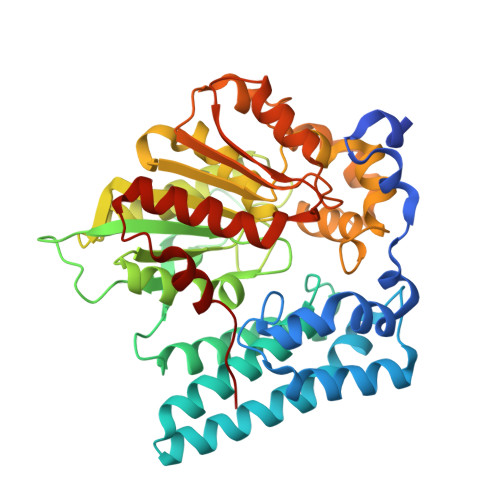
8QD7

Wdyg1p from W. dermatitidis catalyzes polyketide shortening in the biosynthesis of DHN-melanin


Experimental Data Snapshot

  • Method: X-RAY DIFFRACTION
  • Resolution: 1.85 Å
  • R-Value Free: 0.197 
  • R-Value Work: 0.157 
  • R-Value Observed: 0.159 

Starting Model: experimental
View more details

wwPDB Validation   3D Report Full Report


This is version 1.2 of the entry. See complete history


Literature

Polyketide Trimming Shapes Dihydroxynaphthalene-Melanin and Anthraquinone Pigments.

Schmalhofer, M.Vagstad, A.L.Zhou, Q.Bode, H.B.Groll, M.

(2024) Adv Sci (Weinh) 11: e2400184-e2400184

  • DOI: https://doi.org/10.1002/advs.202400184
  • Primary Citation of Related Structures:  
    8QBH, 8QBI, 8QD1, 8QD2, 8QD3, 8QD4, 8QD5, 8QD6, 8QD7, 8QD8, 8QD9, 8QDA, 8QDB

  • PubMed Abstract: 

    Pigments such as anthraquinones (AQs) and melanins are antioxidants, protectants, or virulence factors. AQs from the entomopathogenic bacterium Photorhabdus laumondii are produced by a modular type II polyketide synthase system. A key enzyme involved in AQ biosynthesis is PlAntI, which catalyzes the hydrolysis of the bicyclic-intermediate-loaded acyl carrier protein, polyketide trimming, and assembly of the aromatic AQ scaffold. Here, multiple crystal structures of PlAntI in various conformations and with bound substrate surrogates or inhibitors are reported. Structure-based mutagenesis and activity assays provide experimental insights into the three sequential reaction steps to yield the natural product AQ-256. For comparison, a series of ligand-complex structures of two functionally related hydrolases involved in the biosynthesis of 1,8-dihydroxynaphthalene-melanin in pathogenic fungi is determined. These data provide fundamental insights into the mechanism of polyketide trimming that shapes pigments in pro- and eukaryotes.


  • Organizational Affiliation

    TUM School of Natural Sciences, Department of Bioscience, Centre for Protein Assemblies, Chair of Biochemistry, Technical University of Munich, 85748, Garching, Germany.


Macromolecules
Find similar proteins by:  (by identity cutoff)  |  3D Structure
Entity ID: 1
MoleculeChains Sequence LengthOrganismDetailsImage
Yellowish-green 1-like protein
A, B
431Exophiala dermatitidisMutation(s): 0 
Gene Names: yg1
UniProt
Find proteins for Q6BCB5 (Exophiala dermatitidis)
Explore Q6BCB5 
Go to UniProtKB:  Q6BCB5
Entity Groups  
Sequence Clusters30% Identity50% Identity70% Identity90% Identity95% Identity100% Identity
UniProt GroupQ6BCB5
Sequence Annotations
Expand
  • Reference Sequence
Experimental Data & Validation

Experimental Data

  • Method: X-RAY DIFFRACTION
  • Resolution: 1.85 Å
  • R-Value Free: 0.197 
  • R-Value Work: 0.157 
  • R-Value Observed: 0.159 
  • Space Group: P 21 21 21
Unit Cell:
Length ( Å )Angle ( ˚ )
a = 77.02α = 90
b = 86.62β = 90
c = 115.65γ = 90
Software Package:
Software NamePurpose
REFMACrefinement
XDSdata reduction
XSCALEdata scaling
REFMACphasing

Structure Validation

View Full Validation Report


Entry History & Funding Information

Deposition Data


Funding OrganizationLocationGrant Number
German Research Foundation (DFG)GermanySFB 1309 - 325871075

Revision History  (Full details and data files)

  • Version 1.0: 2024-03-13
    Type: Initial release
  • Version 1.1: 2024-03-27
    Changes: Database references
  • Version 1.2: 2024-06-19
    Changes: Database references