8SQ0 | pdb_00008sq0

Cleaved Ycf1p Dimer in the IFwide-beta conformation


Experimental Data Snapshot

  • Method: ELECTRON MICROSCOPY
  • Resolution: 3.20 Å
  • Aggregation State: PARTICLE 
  • Reconstruction Method: SINGLE PARTICLE 

wwPDB Validation   3D Report Full Report


This is version 1.1 of the entry. See complete history


Literature

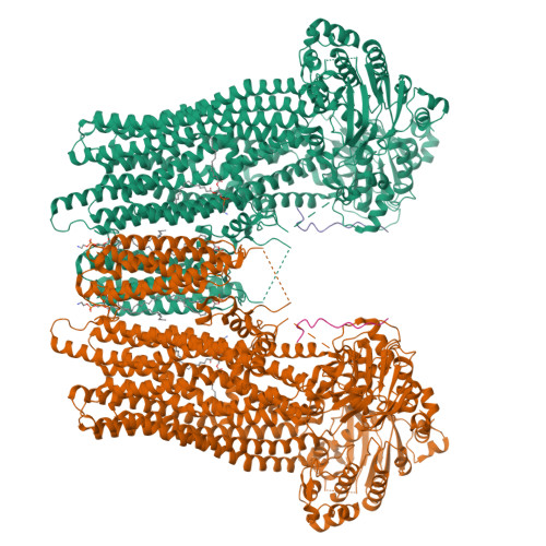
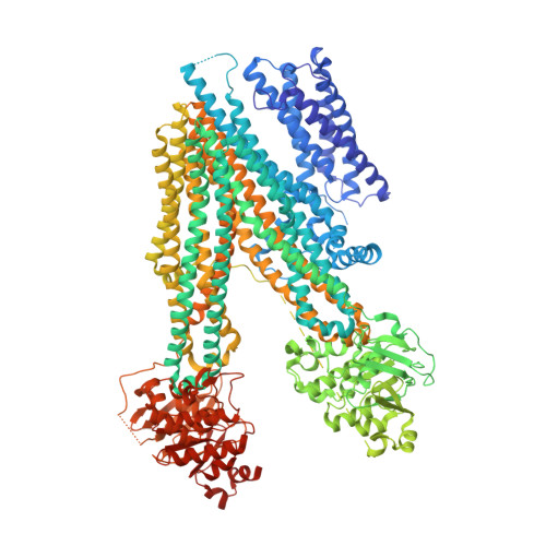
Structure of a dimeric full-length ABC transporter.

Bickers, S.C.Benlekbir, S.Rubinstein, J.L.Kanelis, V.

(2024) Nat Commun 15: 9946-9946

  • DOI: https://doi.org/10.1038/s41467-024-54147-8
  • Primary Citation of Related Structures:  
    8SQ0, 8SQM

  • PubMed Abstract: 

    Activities of ATP binding cassette (ABC) proteins are regulated by multiple mechanisms, including protein interactions, phosphorylation, proteolytic processing, and/or oligomerization of the ABC protein itself. Here we present the structure of yeast cadmium factor 1 (Ycf1p) in its mature form following cleavage by Pep4p protease. Ycf1p, a C subfamily ABC protein (ABCC), is homologue of human multidrug resistance protein 1. Remarkably, a portion of cleaved Ycf1p forms a well-ordered dimer, alongside monomeric particles also present in solution. While numerous other ABC proteins have been proposed to dimerize, no high-resolution structures have been reported. Both phosphorylation of the regulatory (R) region and ATPase activity are lower in the Ycf1p dimer compared to the monomer, indicating that dimerization affects Ycf1p function. The interface between Ycf1p protomers features protein-protein interactions and contains bound lipids, suggesting that lipids stabilize the dimer. The Ycf1p dimer structure may inform the dimerization interfaces of other ABCC dimers.


  • Organizational Affiliation

    Department of Chemistry, University of Toronto, Toronto, ON, Canada.


Macromolecules
Find similar proteins by:  (by identity cutoff)  |  3D Structure
Entity ID: 1
MoleculeChains Sequence LengthOrganismDetailsImage
Metal resistance protein YCF1
A, B
1,537Saccharomyces cerevisiaeMutation(s): 0 
EC: 7.2.2.2 (PDB Primary Data), 7.6.2.3 (PDB Primary Data)
Membrane Entity: Yes 
UniProt
Find proteins for P39109 (Saccharomyces cerevisiae (strain ATCC 204508 / S288c))
Explore P39109 
Go to UniProtKB:  P39109
Entity Groups  
Sequence Clusters30% Identity50% Identity70% Identity90% Identity95% Identity100% Identity
UniProt GroupP39109
Sequence Annotations
Expand
  • Reference Sequence

Find similar proteins by:  Sequence   |   3D Structure  

Entity ID: 2
MoleculeChains Sequence LengthOrganismDetailsImage
Unknown peptide
C, D
13Saccharomyces cerevisiaeMutation(s): 0 
Sequence Annotations
Expand
  • Reference Sequence
Small Molecules
Ligands 3 Unique
IDChains Name / Formula / InChI Key2D Diagram3D Interactions
PTY (Subject of Investigation/LOI)
Query on PTY

Download Ideal Coordinates CCD File 
E [auth A],
H [auth B]
PHOSPHATIDYLETHANOLAMINE
C40 H80 N O8 P
NJGIRBISCGPRPF-KXQOOQHDSA-N
GP7
Query on GP7

Download Ideal Coordinates CCD File 
F [auth A],
I [auth B]
(1R)-2-{[(S)-(2-aminoethoxy)(hydroxy)phosphoryl]oxy}-1-[(pentadecanoyloxy)methyl]ethyl (12E)-hexadeca-9,12-dienoate
C36 H68 N O8 P
NMIBJXZODRRZEA-NTSPIXPOSA-N
LPP
Query on LPP

Download Ideal Coordinates CCD File 
G [auth A],
J [auth B]
2-(HEXADECANOYLOXY)-1-[(PHOSPHONOOXY)METHYL]ETHYL HEXADECANOATE
C35 H69 O8 P
PORPENFLTBBHSG-MGBGTMOVSA-N
Experimental Data & Validation

Experimental Data

  • Method: ELECTRON MICROSCOPY
  • Resolution: 3.20 Å
  • Aggregation State: PARTICLE 
  • Reconstruction Method: SINGLE PARTICLE 

Structure Validation

View Full Validation Report


Entry History & Funding Information

Deposition Data


Funding OrganizationLocationGrant Number
Natural Sciences and Engineering Research Council (NSERC, Canada)CanadaRGPIN-2020-05835

Revision History  (Full details and data files)

  • Version 1.0: 2024-05-08
    Type: Initial release
  • Version 1.1: 2024-11-27
    Changes: Data collection, Database references, Structure summary