8TF0 | pdb_00008tf0

Crystal structure of Grp94 N-terminal domain bound to the purine inhibitor PU-H36


Experimental Data Snapshot

  • Method: X-RAY DIFFRACTION
  • Resolution: 2.79 Å
  • R-Value Free: 
    0.260 (Depositor), 0.260 (DCC) 
  • R-Value Work: 
    0.228 (Depositor), 0.228 (DCC) 
  • R-Value Observed: 
    0.229 (Depositor) 

Starting Model: experimental
View more details

wwPDB Validation   3D Report Full Report


Ligand Structure Quality Assessment 

Created with Raphaël 2.3.0Worse 01 BetterLigand structure goodness of fit to experimental dataBest fitted ZUYClick on this verticalbar to view details

This is version 1.1 of the entry. See complete history


Literature

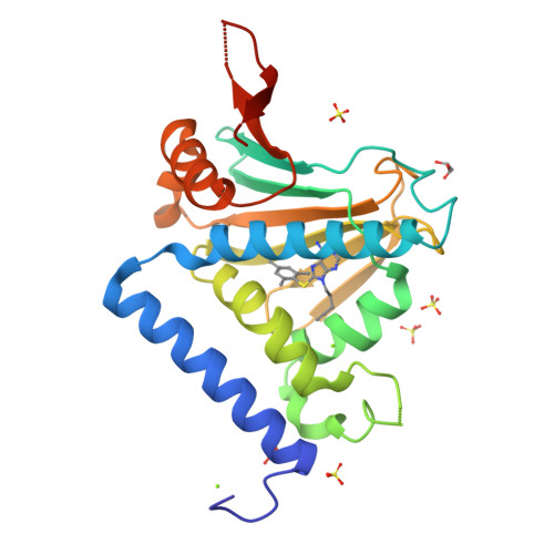
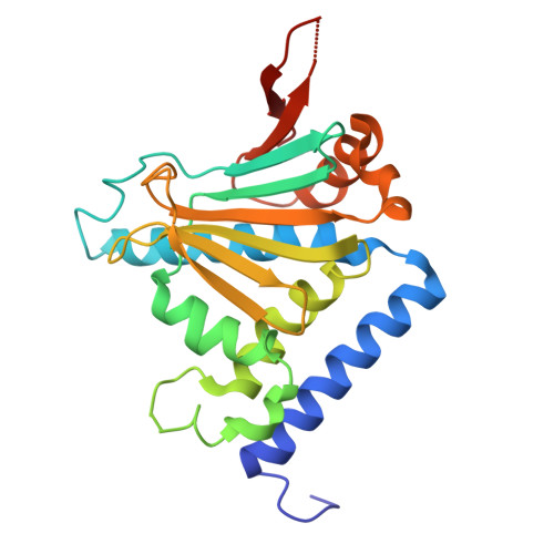
Selective Inhibition of hsp90 Paralogs: Uncovering the Role of Helix 1 in Grp94-Selective Ligand Binding.

Que, N.L.S.Seidler, P.M.Aw, W.J.Chiosis, G.Gewirth, D.T.

(2025) Proteins 93: 654-672

  • DOI: https://doi.org/10.1002/prot.26756
  • Primary Citation of Related Structures:  
    8SBT, 8SSV, 8TF0

  • PubMed Abstract: 

    Grp94 is the endoplasmic reticulum paralog of the hsp90 family of chaperones, which have been targeted for therapeutic intervention via their highly conserved ATP binding sites. The design of paralog-selective inhibitors relies on understanding the protein structural elements that drive higher affinity in selective inhibitors. Here, we determined the structures of Grp94 and Hsp90 in complex with the Grp94-selective inhibitor PU-H36, and of Grp94 with the non-selective inhibitor PU-H71. In Grp94, PU-H36 derives its higher affinity by utilizing Site 2, a Grp94-specific side pocket adjoining the ATP binding cavity, but in Hsp90 PU-H36 occupies Site 1, a side pocket that is accessible in all paralogs with which it makes lower affinity interactions. The structure of Grp94 in complex with PU-H71 shows only Site 1 binding. While changes in the conformation of helices 4 and 5 in the N-terminal domain occur when ligands bind to Site 1 of both Hsp90 and Grp94, large conformational shifts that also involve helix 1 are associated with the engagement of the Site 2 pocket in Grp94 only. Site 2 in Hsp90 is blocked and its helix 1 conformation is insensitive to ligand binding. To understand the role of helix 1 in ligand selectivity, we tested the binding of PU-H36 and other Grp94-selective ligands to chimeric Grp94/Hsp90 constructs. These studies show that helix 1 is the major determinant of selectivity for Site 2 targeted ligands and also influences the rate of ATPase activity in Hsp90 paralogs.


  • Organizational Affiliation

    Hauptman Woodward Medical Research Institute, Buffalo, New York, USA.


Macromolecules
Find similar proteins by:  (by identity cutoff)  |  3D Structure
Entity ID: 1
MoleculeChains Sequence LengthOrganismDetailsImage
EndoplasminA [auth B],
B [auth A],
C,
D
236Canis lupus familiarisMutation(s): 0 
Gene Names: HSP90B1GRP94TRA1
UniProt
Find proteins for P41148 (Canis lupus familiaris)
Explore P41148 
Go to UniProtKB:  P41148
Entity Groups  
Sequence Clusters30% Identity50% Identity70% Identity90% Identity95% Identity100% Identity
UniProt GroupP41148
Sequence Annotations
Expand
  • Reference Sequence
Small Molecules
Ligands 5 Unique
IDChains Name / Formula / InChI Key2D Diagram3D Interactions
ZUY (Subject of Investigation/LOI)
Query on ZUY

Download Ideal Coordinates CCD File 
BA [auth D],
F [auth B],
P [auth A],
Y [auth C]
9-(pent-4-yn-1-yl)-8-[(2,4,6-trimethylphenyl)sulfanyl]-9H-purin-6-amine
C19 H21 N5 S
FFJTXDGAXOSYBK-UHFFFAOYSA-N
SO4
Query on SO4

Download Ideal Coordinates CCD File 
AA [auth C]
IA [auth D]
J [auth B]
JA [auth D]
K [auth B]
SULFATE ION
O4 S
QAOWNCQODCNURD-UHFFFAOYSA-L
GOL
Query on GOL

Download Ideal Coordinates CCD File 
CA [auth D],
G [auth B],
Q [auth A],
R [auth A]
GLYCEROL
C3 H8 O3
PEDCQBHIVMGVHV-UHFFFAOYSA-N
MN
Query on MN

Download Ideal Coordinates CCD File 
DA [auth D]MANGANESE (II) ION
Mn
WAEMQWOKJMHJLA-UHFFFAOYSA-N
MG
Query on MG

Download Ideal Coordinates CCD File 
E [auth B]
EA [auth D]
FA [auth D]
GA [auth D]
H [auth B]
MAGNESIUM ION
Mg
JLVVSXFLKOJNIY-UHFFFAOYSA-N
Experimental Data & Validation

Experimental Data

  • Method: X-RAY DIFFRACTION
  • Resolution: 2.79 Å
  • R-Value Free:  0.260 (Depositor), 0.260 (DCC) 
  • R-Value Work:  0.228 (Depositor), 0.228 (DCC) 
  • R-Value Observed: 0.229 (Depositor) 
Space Group: P 21 21 21
Unit Cell:
Length ( Å )Angle ( ˚ )
a = 96.282α = 90
b = 104.392β = 90
c = 172.284γ = 90
Software Package:
Software NamePurpose
PHENIXrefinement
HKL-2000data reduction
HKL-2000data scaling
MOLREPphasing

Structure Validation

View Full Validation Report


Ligand Structure Quality Assessment 

Created with Raphaël 2.3.0Worse 01 BetterLigand structure goodness of fit to experimental dataBest fitted ZUYClick on this verticalbar to view details

Entry History & Funding Information

Deposition Data


Funding OrganizationLocationGrant Number
National Institutes of Health/National Cancer Institute (NIH/NCI)United StatesP01-CA186866
National Institutes of Health/National Cancer Institute (NIH/NCI)United StatesR01-CA095130

Revision History  (Full details and data files)

  • Version 1.0: 2024-11-06
    Type: Initial release
  • Version 1.1: 2025-02-19
    Changes: Database references