8WM3 | pdb_00008wm3

Cryo-EM structure of ACE2-SIT1 complex with tiagabine


Experimental Data Snapshot

  • Method: ELECTRON MICROSCOPY
  • Resolution: 3.34 Å
  • Aggregation State: PARTICLE 
  • Reconstruction Method: SINGLE PARTICLE 

wwPDB Validation   3D Report Full Report


This is version 1.1 of the entry. See complete history


Literature

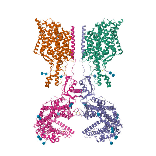
Cryo-EM structure of ACE2-SIT1 in complex with tiagabine.

Broer, A.Hu, Z.Kukulowicz, J.Yadav, A.Zhang, T.Dai, L.Bajda, M.Yan, R.Broer, S.

(2024) J Biological Chem 300: 107687-107687

  • DOI: https://doi.org/10.1016/j.jbc.2024.107687
  • Primary Citation of Related Structures:  
    8WM3

  • PubMed Abstract: 

    The pharmacology of amino acid transporters in the SLC6 family is poorly developed compared to that of the neurotransmitter transporters. To identify new inhibitors of the proline transporter SIT1 (SLC6A20), its expression in Xenopus laevis oocytes was optimized. Trafficking of SIT1 was augmented by co-expression of angiotensin-converting enzyme 2 (ACE2) in oocytes but there was no strict requirement for co-expression of ACE2. A pharmacophore-guided screen identified tiagabine as a potent non-competitive inhibitor of SIT1. To understand its binding mode, we determined the cryo-electron microscopy (cryo-EM) structure of ACE2-SIT1 bound with tiagabine. The inhibitor binds close to the orthosteric proline binding site, but due to its size extends into the cytosolic vestibule. This causes the transporter to adopt an inward-open conformation, in which the intracellular gate is blocked. This study provides the first structural insight into inhibition of SIT1 and generates tools for a better understanding of the ACE2-SIT1 complex. These findings may have significance for SARS-CoV-2 binding to its receptor ACE2 in human lung alveolar cells where SIT1 and ACE2 are functionally expressed.


  • Organizational Affiliation

    Research School of Biology, Australian National University, Canberra, Australia.


Macromolecules
Find similar proteins by:  (by identity cutoff)  |  3D Structure
Entity ID: 1
MoleculeChains Sequence LengthOrganismDetailsImage
Sodium- and chloride-dependent transporter XTRP3
A, C
611Homo sapiensMutation(s): 0 
Gene Names: SLC6A20
Membrane Entity: Yes 
UniProt & NIH Common Fund Data Resources
Find proteins for Q9NP91 (Homo sapiens)
Explore Q9NP91 
Go to UniProtKB:  Q9NP91
PHAROS:  Q9NP91
GTEx:  ENSG00000163817 
Entity Groups  
Sequence Clusters30% Identity50% Identity70% Identity90% Identity95% Identity100% Identity
UniProt GroupQ9NP91
Glycosylation
Glycosylation Sites: 2Go to GlyGen: Q9NP91-1
Sequence Annotations
Expand
  • Reference Sequence
Find similar proteins by:  (by identity cutoff)  |  3D Structure
Entity ID: 2
MoleculeChains Sequence LengthOrganismDetailsImage
Angiotensin-converting enzyme 2
B, D
817Homo sapiensMutation(s): 0 
Gene Names: ACE2UNQ868/PRO1885
EC: 3.4.17.23 (PDB Primary Data), 3.4.17 (PDB Primary Data)
Membrane Entity: Yes 
UniProt & NIH Common Fund Data Resources
Find proteins for Q9BYF1 (Homo sapiens)
Explore Q9BYF1 
Go to UniProtKB:  Q9BYF1
PHAROS:  Q9BYF1
GTEx:  ENSG00000130234 
Entity Groups  
Sequence Clusters30% Identity50% Identity70% Identity90% Identity95% Identity100% Identity
UniProt GroupQ9BYF1
Glycosylation
Glycosylation Sites: 6Go to GlyGen: Q9BYF1-1
Sequence Annotations
Expand
  • Reference Sequence
Oligosaccharides
Experimental Data & Validation

Experimental Data

  • Method: ELECTRON MICROSCOPY
  • Resolution: 3.34 Å
  • Aggregation State: PARTICLE 
  • Reconstruction Method: SINGLE PARTICLE 

Structure Validation

View Full Validation Report


Entry History & Funding Information

Deposition Data


Funding OrganizationLocationGrant Number
National Natural Science Foundation of China (NSFC)China32371267

Revision History  (Full details and data files)

  • Version 1.0: 2024-09-25
    Type: Initial release
  • Version 1.1: 2024-11-20
    Changes: Data collection, Structure summary