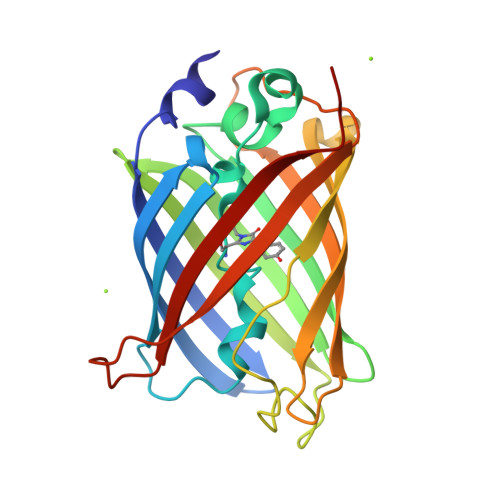
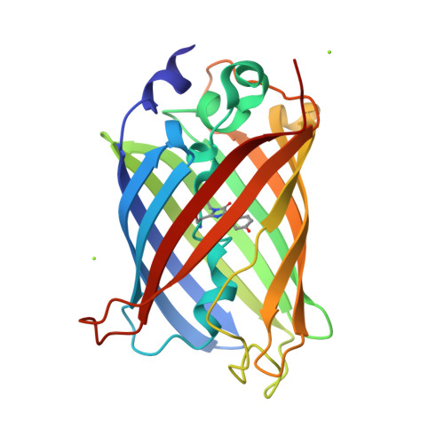
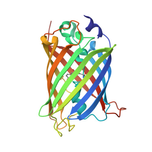
8ZUP

Crystal structure of the F99S/M153T/V163A/T203V/E222Q variant of GFP at pH 8.5


Experimental Data Snapshot

  • Method: X-RAY DIFFRACTION
  • Resolution: 1.20 Å
  • R-Value Free: 0.173 
  • R-Value Work: 0.139 
  • R-Value Observed: 0.141 

Starting Model: experimental
View more details

wwPDB Validation   3D Report Full Report


This is version 1.0 of the entry. See complete history


Literature

Structural characterization of green fluorescent protein in the I-state.

Takeda, R.Tsutsumi, E.Okatsu, K.Fukai, S.Takeda, K.

(2024) Sci Rep 14: 22832-22832

  • DOI: https://doi.org/10.1038/s41598-024-73696-y
  • Primary Citation of Related Structures:  
    8ZUP, 8ZUQ, 8ZUR, 8ZUS, 8ZUT

  • PubMed Abstract: 

    Green fluorescent protein (GFP) is widely utilized as a fluorescent tag in biochemical fields. Whereas the intermediate (I) state has been proposed in the photoreaction cycle in addition to the A and B states, until now the structure of I has only been estimated by computational studies. In this paper, we report the crystal structures of the I stabilizing variants of GFP at high resolutions where respective atoms can be observed separately. Comparison with the structures in the other states highlights the structural feature of the I state. The side chain of one of the substituted residues, Val203, adopts the gauche- conformation observed for Thr203 in the A state, which is different from the B state. On the other hand, His148 interacts with the chromophore by ordinary hydrogen bonding with a distance of 2.85 Å, while the weaker interaction by longer distances is observed in the A state. Therefore, it was indicated that it is possible to distinguish three states A, B and I by the two hydrogen bond distances Oγ-Thr203···Oη-chromophore and Nδ1-His148···Oη-chromophore. We discuss the characteristics of the I intermediate of wild-type GFP on the bases of the structure estimated from the variant structures by quantum chemical calculations.


  • Organizational Affiliation

    Department of Chemistry, Graduate School of Science, Kyoto University, Sakyo-ku, Kyoto, 606-8502, Japan.


Macromolecules
Find similar proteins by:  (by identity cutoff)  |  3D Structure
Entity ID: 1
MoleculeChains Sequence LengthOrganismDetailsImage
Green fluorescent protein228Aequorea victoriaMutation(s): 6 
Gene Names: GFP
UniProt
Find proteins for P42212 (Aequorea victoria)
Explore P42212 
Go to UniProtKB:  P42212
Entity Groups  
Sequence Clusters30% Identity50% Identity70% Identity90% Identity95% Identity100% Identity
UniProt GroupP42212
Sequence Annotations
Expand
  • Reference Sequence
Small Molecules
Modified Residues  1 Unique
IDChains TypeFormula2D DiagramParent
GYS
Query on GYS
A
L-PEPTIDE LINKINGC14 H15 N3 O5SER, TYR, GLY
Experimental Data & Validation

Experimental Data

  • Method: X-RAY DIFFRACTION
  • Resolution: 1.20 Å
  • R-Value Free: 0.173 
  • R-Value Work: 0.139 
  • R-Value Observed: 0.141 
  • Space Group: P 21 21 21
Unit Cell:
Length ( Å )Angle ( ˚ )
a = 50.696α = 90
b = 62.273β = 90
c = 68.429γ = 90
Software Package:
Software NamePurpose
PHENIXrefinement
XDSdata reduction
XDSdata scaling
PHENIXphasing

Structure Validation

View Full Validation Report


Entry History & Funding Information

Deposition Data


Funding OrganizationLocationGrant Number
Takeda Science FoundationJapan--
Gimpu fund from Kyoto UniversityJapan--

Revision History  (Full details and data files)

  • Version 1.0: 2024-11-06
    Type: Initial release