RCSB PDB - 9AT6: Crystal structure of SARS-CoV-2 3CL protease in complex with a methylbicyclo[2.2.1]heptene 2-pyrrolidone inhibitor

 9AT6

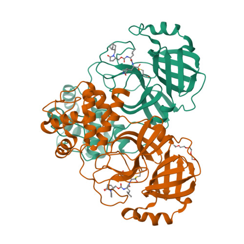
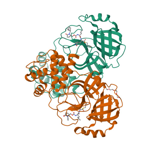
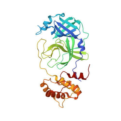
Crystal structure of SARS-CoV-2 3CL protease in complex with a methylbicyclo[2.2.1]heptene 2-pyrrolidone inhibitor


Experimental Data Snapshot

  • Method: X-RAY DIFFRACTION
  • Resolution: 1.40 Å
  • R-Value Free: 0.170 
  • R-Value Work: 0.145 
  • R-Value Observed: 0.146 

Starting Model: experimental
View more details

wwPDB Validation   3D Report Full Report


Ligand Structure Quality Assessment 

Created with Raphaël 2.3.0Worse 01 BetterLigand structure goodness of fit to experimental dataBest fitted A1AGRClick on this verticalbar to view detailsBest fitted A1AGQClick on this verticalbar to view details

This is version 1.2 of the entry. See complete history


Literature

Structure-Guided Design of Potent Coronavirus Inhibitors with a 2-Pyrrolidone Scaffold: Biochemical, Crystallographic, and Virological Studies.

Dampalla, C.S.Kim, Y.Zabiegala, A.Howard, D.J.Nguyen, H.N.Madden, T.K.Thurman, H.A.Cooper, A.Liu, L.Battaile, K.P.Lovell, S.Chang, K.O.Groutas, W.C.

(2024) J Med Chem 67: 11937-11956

  • DOI: https://doi.org/10.1021/acs.jmedchem.4c00551
  • Primary Citation of Related Structures:  
    9ASV, 9ASW, 9ASY, 9ASZ, 9AT0, 9AT1, 9AT3, 9AT4, 9AT5, 9AT6, 9AT7, 9ATA, 9ATD, 9ATE, 9ATF, 9ATG, 9ATH, 9ATI, 9ATJ, 9ATS, 9ATT

  • PubMed Abstract: 

    Zoonotic coronaviruses are known to produce severe infections in humans and have been the cause of significant morbidity and mortality worldwide. SARS-CoV-2 was the largest and latest contributor of fatal cases, even though MERS-CoV has the highest case-fatality ratio among zoonotic coronaviruses. These infections pose a high risk to public health worldwide warranting efforts for the expeditious discovery of antivirals. Hence, we hereby describe a novel series of inhibitors of coronavirus 3CL pro embodying an N -substituted 2-pyrrolidone scaffold envisaged to exploit favorable interactions with the S3-S4 subsites and connected to an invariant Leu-Gln P 2-P1 recognition element. Several inhibitors showed nanomolar antiviral activity in enzyme and cell-based assays, with no significant cytotoxicity. High-resolution crystal structures of inhibitors bound to the 3CL pro were determined to probe and identify the molecular determinants associated with binding, to inform the structure-guided optimization of the inhibitors, and to confirm the mechanism of action of the inhibitors.


  • Organizational Affiliation

    Department of Chemistry and Biochemistry, Wichita State University, Wichita, Kansas 67260, United States.


Macromolecules
Find similar proteins by:  (by identity cutoff)  |  3D Structure
Entity ID: 1
MoleculeChains Sequence LengthOrganismDetailsImage
3C-like proteinase nsp5
A, B
308Severe acute respiratory syndrome coronavirus 2Mutation(s): 0 
Gene Names: rep1a-1b
EC: 3.4.22.69
UniProt
Find proteins for P0DTD1 (Severe acute respiratory syndrome coronavirus 2)
Explore P0DTD1 
Go to UniProtKB:  P0DTD1
Entity Groups  
Sequence Clusters30% Identity50% Identity70% Identity90% Identity95% Identity100% Identity
UniProt GroupP0DTD1
Sequence Annotations
Expand
  • Reference Sequence
Small Molecules
Ligands 3 Unique
IDChains Name / Formula / InChI Key2D Diagram3D Interactions
A1AGR
Query on A1AGR

Download Ideal Coordinates CCD File 
D [auth A],
E [auth B]
(1S,2S)-2-{[N-({[(2S)-1-{[(1R,2S,4R)-bicyclo[2.2.1]hept-5-en-2-yl]methyl}-5-oxopyrrolidin-2-yl]methoxy}carbonyl)-L-leucyl]amino}-1-hydroxy-3-[(3S)-2-oxopyrrolidin-3-yl]propane-1-sulfonic acid
C27 H42 N4 O9 S
DZWIJESAJPXJIB-XTXIHYAPSA-N
A1AGQ
Query on A1AGQ

Download Ideal Coordinates CCD File 
C [auth A],
F [auth B]
(1R,2S)-2-{[N-({[(2S)-1-{[(1R,2S,4R)-bicyclo[2.2.1]hept-5-en-2-yl]methyl}-5-oxopyrrolidin-2-yl]methoxy}carbonyl)-L-leucyl]amino}-1-hydroxy-3-[(3S)-2-oxopyrrolidin-3-yl]propane-1-sulfonic acid
C27 H42 N4 O9 S
DZWIJESAJPXJIB-AQOOIRESSA-N
PG4
Query on PG4

Download Ideal Coordinates CCD File 
G [auth B]TETRAETHYLENE GLYCOL
C8 H18 O5
UWHCKJMYHZGTIT-UHFFFAOYSA-N
Experimental Data & Validation

Experimental Data

  • Method: X-RAY DIFFRACTION
  • Resolution: 1.40 Å
  • R-Value Free: 0.170 
  • R-Value Work: 0.145 
  • R-Value Observed: 0.146 
  • Space Group: P 1 21 1
Unit Cell:
Length ( Å )Angle ( ˚ )
a = 55.237α = 90
b = 98.287β = 107.89
c = 58.911γ = 90
Software Package:
Software NamePurpose
PHENIXrefinement
Aimlessdata scaling
XDSdata reduction
PHASERphasing

Structure Validation

View Full Validation Report


Ligand Structure Quality Assessment 

Created with Raphaël 2.3.0Worse 01 BetterLigand structure goodness of fit to experimental dataBest fitted A1AGRClick on this verticalbar to view detailsBest fitted A1AGQClick on this verticalbar to view details

Entry History & Funding Information

Deposition Data


Funding OrganizationLocationGrant Number
National Institutes of Health/National Institute Of Allergy and Infectious Diseases (NIH/NIAID)United StatesR01AI161085
National Institutes of Health/Office of the DirectorUnited StatesS10OD030394

Revision History  (Full details and data files)

  • Version 1.0: 2024-07-10
    Type: Initial release
  • Version 1.1: 2024-08-07
    Changes: Database references
  • Version 1.2: 2024-10-09
    Changes: Structure summary