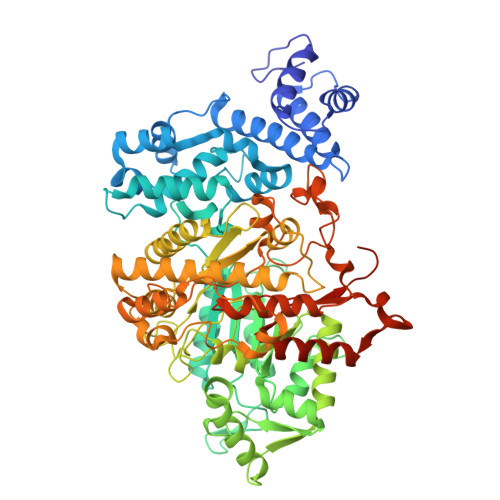
9DB2

Class Ia ribonucleotide reductase with mechanism-based inhibitor N3CDP


Experimental Data Snapshot

  • Method: ELECTRON MICROSCOPY
  • Resolution: 2.60 Å
  • Aggregation State: PARTICLE 
  • Reconstruction Method: SINGLE PARTICLE 

wwPDB Validation   3D Report Full Report


This is version 1.1 of the entry. See complete history


Literature

2.6- angstrom resolution cryo-EM structure of a class Ia ribonucleotide reductase trapped with mechanism-based inhibitor N 3 CDP.

Westmoreland, D.E.Feliciano, P.R.Kang, G.Cui, C.Kim, A.Stubbe, J.Nocera, D.G.Drennan, C.L.

(2024) Proc Natl Acad Sci U S A 121: e2417157121-e2417157121

  • DOI: https://doi.org/10.1073/pnas.2417157121
  • Primary Citation of Related Structures:  
    9DB2

  • PubMed Abstract: 

    Ribonucleotide reductases (RNRs) reduce ribonucleotides to deoxyribonucleotides using radical-based chemistry. For class Ia RNRs, the radical species is stored in a separate subunit (β2) from the subunit housing the active site (α2), requiring the formation of a short-lived α2β2 complex and long-range radical transfer (RT). RT occurs via proton-coupled electron transfer (PCET) over a long distance (~32-Å) and involves the formation and decay of multiple amino acid radical species. Here, we use cryogenic electron microscopy and a mechanism-based inhibitor 2'-azido-2'-deoxycytidine-5'-diphosphate (N 3 CDP) to trap a wild-type α2β2 complex of Escherichia coli class Ia RNR. We find that one α subunit has turned over and that the other is trapped, bound to β in a midturnover state. Instead of N 3 CDP in the active site, forward RT has resulted in N 2 loss, migration of the third nitrogen from the ribose C2' to C3' positions, and attachment of this nitrogen to the sulfur of cysteine-225. In this study, an inhibitor has been visualized as an adduct to an RNR. Additionally, this structure reveals the positions of PCET residues following forward RT, complementing the previous structure that depicted a preturnover PCET pathway and suggesting how PCET is gated at the α-β interface. This N 3 CDP-trapped structure is also of sufficient resolution (2.6 Å) to visualize water molecules, allowing us to evaluate the proposal that water molecules are proton acceptors and donors as part of the PCET process.


  • Organizational Affiliation

    Department of Biology, Massachusetts Institute of Technology, Cambridge, MA 02139.


Macromolecules
Find similar proteins by:  (by identity cutoff)  |  3D Structure
Entity ID: 1
MoleculeChains Sequence LengthOrganismDetailsImage
Ribonucleoside-diphosphate reductase 1 subunit alpha
A, B
761Escherichia coliMutation(s): 0 
Gene Names: nrdAdnaFb2234JW2228
EC: 1.17.4.1
UniProt
Find proteins for P00452 (Escherichia coli (strain K12))
Explore P00452 
Go to UniProtKB:  P00452
Entity Groups  
Sequence Clusters30% Identity50% Identity70% Identity90% Identity95% Identity100% Identity
UniProt GroupP00452
Sequence Annotations
Expand
  • Reference Sequence
Find similar proteins by:  (by identity cutoff)  |  3D Structure
Entity ID: 2
MoleculeChains Sequence LengthOrganismDetailsImage
Ribonucleoside-diphosphate reductase 1 subunit beta
C, D
376Escherichia coliMutation(s): 0 
Gene Names: nrdBftsBb2235JW2229
EC: 1.17.4.1
UniProt
Find proteins for P69924 (Escherichia coli (strain K12))
Explore P69924 
Go to UniProtKB:  P69924
Entity Groups  
Sequence Clusters30% Identity50% Identity70% Identity90% Identity95% Identity100% Identity
UniProt GroupP69924
Sequence Annotations
Expand
  • Reference Sequence
Small Molecules
Ligands 6 Unique
IDChains Name / Formula / InChI Key2D Diagram3D Interactions
ATP (Subject of Investigation/LOI)
Query on ATP

Download Ideal Coordinates CCD File 
G [auth A],
H [auth A],
K [auth B],
L [auth B]
ADENOSINE-5'-TRIPHOSPHATE
C10 H16 N5 O13 P3
ZKHQWZAMYRWXGA-KQYNXXCUSA-N
DTP (Subject of Investigation/LOI)
Query on DTP

Download Ideal Coordinates CCD File 
E [auth A],
M [auth B]
2'-DEOXYADENOSINE 5'-TRIPHOSPHATE
C10 H16 N5 O12 P3
SUYVUBYJARFZHO-RRKCRQDMSA-N
A1A3L (Subject of Investigation/LOI)
Query on A1A3L

Download Ideal Coordinates CCD File 
P [auth B]4-amino-1-{(3xi)-3-C-amino-2-deoxy-5-O-[(S)-hydroxy(phosphonooxy)phosphoryl]-beta-D-threo-pentofuranosyl}pyrimidin-2(1H)-one
C9 H16 N4 O10 P2
XAPJNFNJUSYHIU-SLBFFKMLSA-N
FEO (Subject of Investigation/LOI)
Query on FEO

Download Ideal Coordinates CCD File 
Q [auth C],
R [auth D]
MU-OXO-DIIRON
Fe2 O
NPMYUMBHPJGBFA-UHFFFAOYSA-N
MG
Query on MG

Download Ideal Coordinates CCD File 
F [auth A],
I [auth A],
N [auth B],
O [auth B]
MAGNESIUM ION
Mg
JLVVSXFLKOJNIY-UHFFFAOYSA-N
UNL (Subject of Investigation/LOI)
Query on UNL

Download Ideal Coordinates CCD File 
J [auth A]Unknown ligand
JLVVSXFLKOJNIY-UHFFFAOYSA-N
Experimental Data & Validation

Experimental Data

  • Method: ELECTRON MICROSCOPY
  • Resolution: 2.60 Å
  • Aggregation State: PARTICLE 
  • Reconstruction Method: SINGLE PARTICLE 
EM Software:
TaskSoftware PackageVersion
MODEL REFINEMENTPHENIX1.20.1_4487:

Structure Validation

View Full Validation Report


Entry History & Funding Information

Deposition Data


Funding OrganizationLocationGrant Number
National Institutes of Health/National Institute of General Medical Sciences (NIH/NIGMS)United States1R35GM126982-01
National Institutes of Health/National Institute of General Medical Sciences (NIH/NIGMS)United StatesGM047274
National Institutes of Health/National Institute of General Medical Sciences (NIH/NIGMS)United StatesGM29595
National Institutes of Health/National Institute of General Medical Sciences (NIH/NIGMS)United States1F32GM145072-01

Revision History  (Full details and data files)

  • Version 1.0: 2024-11-06
    Type: Initial release
  • Version 1.1: 2024-11-27
    Changes: Data collection