9G13 | pdb_00009g13

VHH H3-2 in complex with Tau C-terminal peptide


Experimental Data Snapshot

  • Method: X-RAY DIFFRACTION
  • Resolution: 1.80 Å
  • R-Value Free: 
    0.231 (Depositor), 0.240 (DCC) 
  • R-Value Work: 
    0.172 (Depositor), 0.180 (DCC) 

Starting Model: experimental
View more details

wwPDB Validation   3D Report Full Report


This is version 1.2 of the entry. See complete history


Literature

Inhibition of tau neuronal internalization using anti-tau single domain antibodies.

Danis, C.Dupre, E.Bouillet, T.Denechaud, M.Lefebvre, C.Nguyen, M.Mortelecque, J.Cantrelle, F.X.Rain, J.C.Hanoulle, X.Colin, M.Buee, L.Landrieu, I.

(2025) Nat Commun 16: 3162-3162

  • DOI: https://doi.org/10.1038/s41467-025-58383-4
  • Primary Citation of Related Structures:  
    9G13

  • PubMed Abstract: 

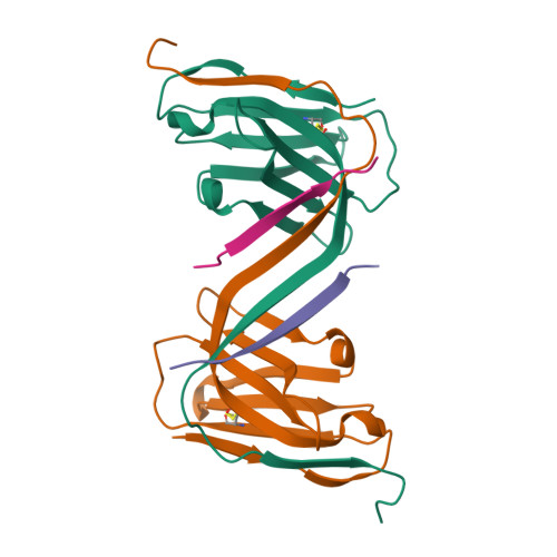
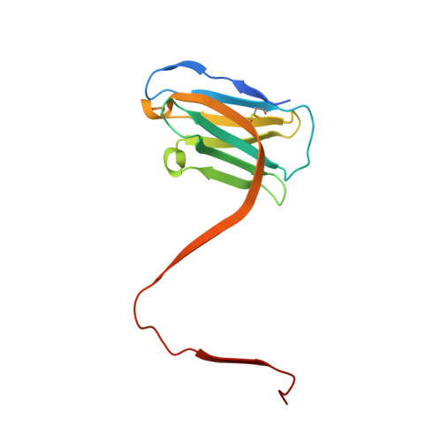
    In Alzheimer's disease, tau pathology spreads across brain regions as the disease progresses. Intracellular tau can be released and taken up by nearby neurons. We evaluated single domain anti-tau antibodies, also called VHHs, as inhibitors of tau internalization. We identified three VHH inhibitors of tau uptake: A31, H3-2, and Z70 mut1 . These VHHs compete with the membrane protein LRP1, a major receptor mediating neuronal uptake of tau. A31 and Z70 mut1 bind to microtubule binding domain repeats, which are involved in the interaction with LRP1. VHH H3-2 is the only VHH from our library that reduces the internalization of both monomeric tau and tau fibrils. VHH H3-2 binds a C-terminal tau epitope with high affinity. Its three-dimensional structure in complex with a tau peptide reveals a unique binding mode as a VHH-swapped dimer. These anti-tau VHHs are interesting tools to study tau prion-like propagation in tauopathies and potentially develop novel biotherapies.


  • Organizational Affiliation

    CNRS EMR9002 - BSI - Integrative Structural Biology, Lille, France. clement.danis@inserm.fr.


Macromolecules
Find similar proteins by:  (by identity cutoff)  |  3D Structure
Entity ID: 1
MoleculeChains Sequence LengthOrganismDetailsImage
VHH H3-2A,
C [auth E],
E [auth C],
G
132Lama glamaMutation(s): 0 
Entity Groups  
Sequence Clusters30% Identity50% Identity70% Identity90% Identity95% Identity100% Identity
Sequence Annotations
Expand
  • Reference Sequence

Find similar proteins by:  Sequence   |   3D Structure  

Entity ID: 2
MoleculeChains Sequence LengthOrganismDetailsImage
Isoform Tau-F of Microtubule-associated protein tauB,
D [auth F],
F [auth D],
H
13Homo sapiensMutation(s): 0 
UniProt & NIH Common Fund Data Resources
Find proteins for P10636 (Homo sapiens)
Explore P10636 
Go to UniProtKB:  P10636
PHAROS:  P10636
GTEx:  ENSG00000186868 
Entity Groups  
Sequence Clusters30% Identity50% Identity70% Identity90% Identity95% Identity100% Identity
UniProt GroupP10636
Sequence Annotations
Expand
  • Reference Sequence
Experimental Data & Validation

Experimental Data

  • Method: X-RAY DIFFRACTION
  • Resolution: 1.80 Å
  • R-Value Free:  0.231 (Depositor), 0.240 (DCC) 
  • R-Value Work:  0.172 (Depositor), 0.180 (DCC) 
Space Group: P 21 21 21
Unit Cell:
Length ( Å )Angle ( ˚ )
a = 70.394α = 90
b = 76.346β = 90
c = 95.565γ = 90
Software Package:
Software NamePurpose
REFMACrefinement
Cootmodel building
Aimlessdata scaling
XDSdata reduction
pointlessdata scaling
MOLREPphasing

Structure Validation

View Full Validation Report


Entry History & Funding Information

Deposition Data


Funding OrganizationLocationGrant Number
Agence Nationale de la Recherche (ANR)FranceANR-18-CE44-0016
Agence Nationale de la Recherche (ANR)FranceANR-11-LABX-01

Revision History  (Full details and data files)

  • Version 1.0: 2024-07-31
    Type: Initial release
  • Version 1.1: 2024-11-20
    Changes: Structure summary
  • Version 1.2: 2025-04-09
    Changes: Database references