4BEM | pdb_00004bem

Crystal structure of the F-type ATP synthase c-ring from Acetobacterium woodii.


Experimental Data Snapshot

  • Method: X-RAY DIFFRACTION
  • Resolution: 2.10 Å
  • R-Value Free: 
    0.220 (Depositor), 0.220 (DCC) 
  • R-Value Work: 
    0.182 (Depositor), 0.190 (DCC) 
  • R-Value Observed: 
    0.184 (Depositor) 

Starting Model: experimental
View more details

wwPDB Validation   3D Report Full Report


Ligand Structure Quality Assessment 

Created with Raphaël 2.3.0Worse 01 BetterLigand structure goodness of fit to experimental dataBest fitted HTGClick on this verticalbar to view details

This is version 1.5 of the entry. See complete history


Literature

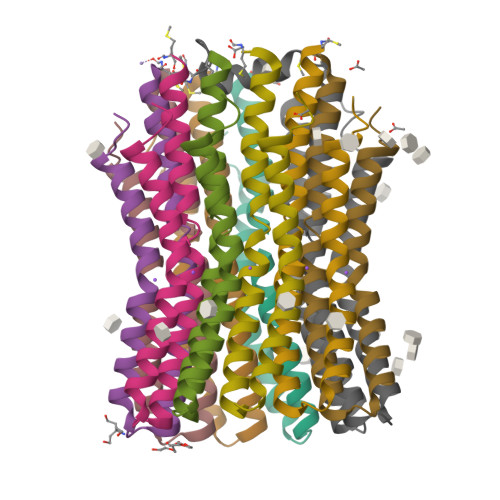
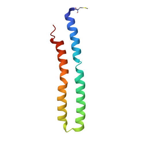
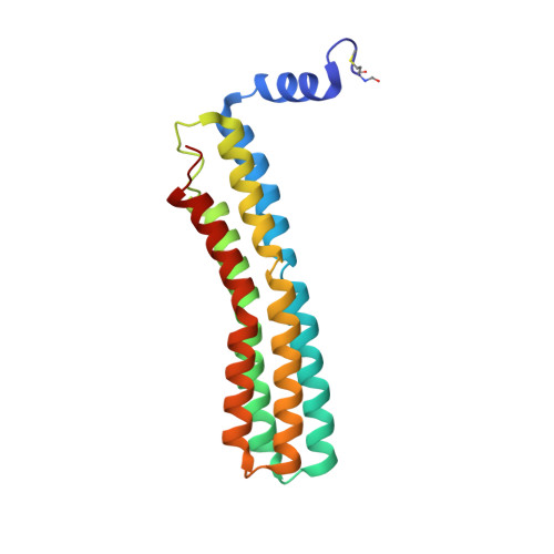
High-Resolution Structure and Mechanism of an F/V-Hybrid Rotor Ring in a Na+-Coupled ATP Synthase

Matthies, D.Zhou, W.Klyszejko, A.L.Anselmi, C.Yildiz, O.Brandt, K.Muller, V.Faraldo-Gomez, J.D.Meier, T.

(2014) Nat Commun 5: 5286

  • DOI: https://doi.org/10.1038/ncomms6286
  • Primary Citation of Related Structures:  
    4BEM

  • PubMed Abstract: 

    All rotary ATPases catalyse the interconversion of ATP and ADP-Pi through a mechanism that is coupled to the transmembrane flow of H(+) or Na(+). Physiologically, however, F/A-type enzymes specialize in ATP synthesis driven by downhill ion diffusion, while eukaryotic V-type ATPases function as ion pumps. To begin to rationalize the molecular basis for this functional differentiation, we solved the crystal structure of the Na(+)-driven membrane rotor of the Acetobacterium woodii ATP synthase, at 2.1 Å resolution. Unlike known structures, this rotor ring is a 9:1 heteromer of F- and V-type c-subunits and therefore features a hybrid configuration of ion-binding sites along its circumference. Molecular and kinetic simulations are used to dissect the mechanisms of Na(+) recognition and rotation of this c-ring, and to explain the functional implications of the V-type c-subunit. These structural and mechanistic insights indicate an evolutionary path between synthases and pumps involving adaptations in the rotor ring.


  • Organizational Affiliation

    Department of Structural Biology, Max Planck Institute of Biophysics, Max-von-Laue-Str. 3, 60438 Frankfurt am Main, Germany.


Macromolecules
Find similar proteins by:  (by identity cutoff)  |  3D Structure
Entity ID: 1
MoleculeChains Sequence LengthOrganismDetailsImage
F1FO ATPASE C2 SUBUNIT
A, B, C, D, E
82Acetobacterium woodii DSM 1030Mutation(s): 0 
Membrane Entity: Yes 
UniProt
Find proteins for H6LFT2 (Acetobacterium woodii (strain ATCC 29683 / DSM 1030 / JCM 2381 / KCTC 1655 / WB1))
Explore H6LFT2 
Go to UniProtKB:  H6LFT2
Entity Groups  
Sequence Clusters30% Identity50% Identity70% Identity90% Identity95% Identity100% Identity
UniProt GroupH6LFT2
Sequence Annotations
Expand
  • Reference Sequence
Find similar proteins by:  (by identity cutoff)  |  3D Structure
Entity ID: 2
MoleculeChains Sequence LengthOrganismDetailsImage
F1FO ATPASE C1 SUBUNIT182Acetobacterium woodii DSM 1030Mutation(s): 0 
Membrane Entity: Yes 
UniProt
Find proteins for H6LFT0 (Acetobacterium woodii (strain ATCC 29683 / DSM 1030 / JCM 2381 / KCTC 1655 / WB1))
Explore H6LFT0 
Go to UniProtKB:  H6LFT0
Entity Groups  
Sequence Clusters30% Identity50% Identity70% Identity90% Identity95% Identity100% Identity
UniProt GroupH6LFT0
Sequence Annotations
Expand
  • Reference Sequence
Small Molecules
Ligands 6 Unique
IDChains Name / Formula / InChI Key2D Diagram3D Interactions
HTG
Query on HTG

Download Ideal Coordinates CCD File 
AA [auth G]
BA [auth G]
CA [auth G]
EA [auth H]
FA [auth H]
heptyl 1-thio-beta-D-glucopyranoside
C13 H26 O5 S
HPEGNLMTTNTJSP-LBELIVKGSA-N
P6G
Query on P6G

Download Ideal Coordinates CCD File 
S [auth E]HEXAETHYLENE GLYCOL
C12 H26 O7
IIRDTKBZINWQAW-UHFFFAOYSA-N
TAM
Query on TAM

Download Ideal Coordinates CCD File 
P [auth C]TRIS(HYDROXYETHYL)AMINOMETHANE
C7 H17 N O3
GKODZWOPPOTFGA-UHFFFAOYSA-N
ACT
Query on ACT

Download Ideal Coordinates CCD File 
HA [auth I],
IA [auth I],
X [auth F],
Z [auth G]
ACETATE ION
C2 H3 O2
QTBSBXVTEAMEQO-UHFFFAOYSA-M
MN
Query on MN

Download Ideal Coordinates CCD File 
O [auth C]MANGANESE (II) ION
Mn
WAEMQWOKJMHJLA-UHFFFAOYSA-N
NA
Query on NA

Download Ideal Coordinates CCD File 
DA [auth H]
GA [auth I]
K [auth A]
L [auth B]
N [auth C]
SODIUM ION
Na
FKNQFGJONOIPTF-UHFFFAOYSA-N
Modified Residues  1 Unique
IDChains TypeFormula2D DiagramParent
FME
Query on FME
A, B, C, D, E
L-PEPTIDE LINKINGC6 H11 N O3 SMET
Experimental Data & Validation

Experimental Data

  • Method: X-RAY DIFFRACTION
  • Resolution: 2.10 Å
  • R-Value Free:  0.220 (Depositor), 0.220 (DCC) 
  • R-Value Work:  0.182 (Depositor), 0.190 (DCC) 
  • R-Value Observed: 0.184 (Depositor) 
Space Group: P 43 21 2
Unit Cell:
Length ( Å )Angle ( ˚ )
a = 121.17α = 90
b = 121.17β = 90
c = 150.57γ = 90
Software Package:
Software NamePurpose
PHENIXrefinement
XDSdata reduction
XSCALEdata scaling
PHASERphasing

Structure Validation

View Full Validation Report


Ligand Structure Quality Assessment 

Created with Raphaël 2.3.0Worse 01 BetterLigand structure goodness of fit to experimental dataBest fitted HTGClick on this verticalbar to view details

Entry History 

Deposition Data

Revision History  (Full details and data files)

  • Version 1.0: 2014-03-26
    Type: Initial release
  • Version 1.1: 2014-10-01
    Changes: Database references
  • Version 1.2: 2014-11-19
    Changes: Database references
  • Version 1.3: 2020-07-29
    Type: Remediation
    Reason: Carbohydrate remediation
    Changes: Derived calculations, Other, Structure summary
  • Version 1.4: 2023-12-20
    Changes: Data collection, Database references, Refinement description, Structure summary
  • Version 1.5: 2024-10-23
    Changes: Structure summary