5FOR | pdb_00005for

Cryptic TIR


Experimental Data Snapshot

  • Method: X-RAY DIFFRACTION
  • Resolution: 2.50 Å
  • R-Value Free: 
    0.244 (Depositor), 0.250 (DCC) 
  • R-Value Work: 
    0.188 (Depositor), 0.200 (DCC) 
  • R-Value Observed: 
    0.191 (Depositor) 

wwPDB Validation   3D Report Full Report


Ligand Structure Quality Assessment 

Created with Raphaël 2.3.0Worse 01 BetterLigand structure goodness of fit to experimental dataBest fitted I3CClick on this verticalbar to view details

This is version 1.2 of the entry. See complete history


Literature

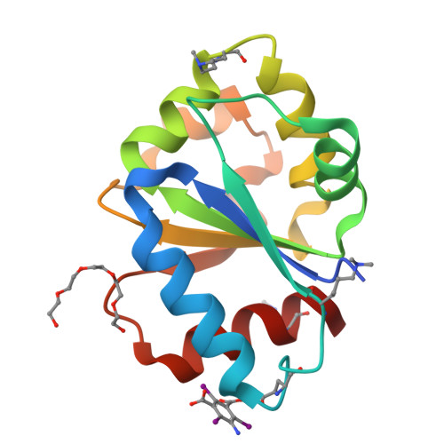
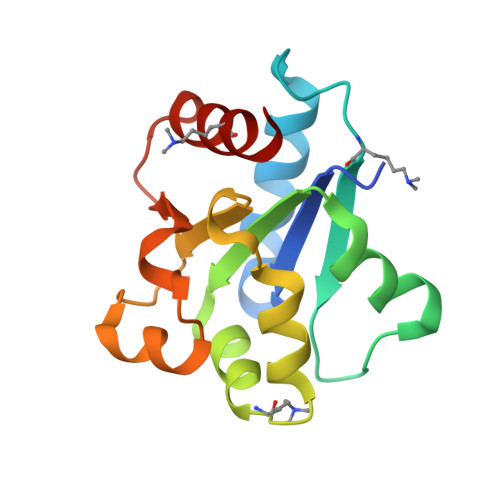
Structure of the Toll/Interleukin-1 Receptor (TIR) Domain of the B-cell Adaptor That Links Phosphoinositide Metabolism with the Negative Regulation of the Toll-like Receptor (TLR) Signalosome.

Halabi, S.Sekine, E.Verstak, B.Gay, N.J.Moncrieffe, M.C.

(2017) J Biological Chem 292: 652-660

  • DOI: https://doi.org/10.1074/jbc.M116.761528
  • Primary Citation of Related Structures:  
    5FOR

  • PubMed Abstract: 

    Ligand binding to Toll-like receptors (TLRs) results in dimerization of their cytosolic Toll/interleukin-1 receptor (TIR) domains and recruitment of post-receptor signal transducers into a complex signalosome. TLR activation leads to the production of transcription factors and pro-inflammatory molecules and the activation of phosphoinositide 3-kinases (PI3K) in a process that requires the multimodular B-cell adaptor for phosphoinositide 3-kinase (BCAP). BCAP has a sequence previously proposed as a "cryptic" TIR domain. Here, we present the structure of the N-terminal region of human BCAP and show that it possesses a canonical TIR fold. Dimeric BCAP associates with the TIR domains of TLR2/4 and MAL/TIRAP, suggesting that it is recruited to the TLR signalosome by multitypic TIR-TIR interactions. BCAP also interacts with the p85 subunit of PI3K and phospholipase Cγ, enzymes that deplete plasma membrane phosphatidylinositol 4,5-bisphosphate (PIP2), and these interactions provide a molecular explanation for BCAP-mediated down-regulation of inflammatory signaling.


  • Organizational Affiliation

    From the Department of Biochemistry, Cambridge University, Cambridge CB2 1GA, United Kingdom.


Macromolecules
Find similar proteins by:  (by identity cutoff)  |  3D Structure
Entity ID: 1
MoleculeChains Sequence LengthOrganismDetailsImage
PHOSPHOINOSITIDE 3-KINASE ADAPTER PROTEIN 1139Homo sapiensMutation(s): 0 
UniProt & NIH Common Fund Data Resources
Find proteins for Q6ZUJ8 (Homo sapiens)
Explore Q6ZUJ8 
Go to UniProtKB:  Q6ZUJ8
PHAROS:  Q6ZUJ8
GTEx:  ENSG00000155629 
Entity Groups  
Sequence Clusters30% Identity50% Identity70% Identity90% Identity95% Identity100% Identity
UniProt GroupQ6ZUJ8
Sequence Annotations
Expand
  • Reference Sequence
Small Molecules
Ligands 2 Unique
IDChains Name / Formula / InChI Key2D Diagram3D Interactions
I3C
Query on I3C

Download Ideal Coordinates CCD File 
C [auth A]5-amino-2,4,6-triiodobenzene-1,3-dicarboxylic acid
C8 H4 I3 N O4
JEZJSNULLBSYHV-UHFFFAOYSA-N
1PE
Query on 1PE

Download Ideal Coordinates CCD File 
B [auth A]PENTAETHYLENE GLYCOL
C10 H22 O6
JLFNLZLINWHATN-UHFFFAOYSA-N
Modified Residues  1 Unique
IDChains TypeFormula2D DiagramParent
MLY
Query on MLY
A
L-PEPTIDE LINKINGC8 H18 N2 O2LYS
Experimental Data & Validation

Experimental Data

  • Method: X-RAY DIFFRACTION
  • Resolution: 2.50 Å
  • R-Value Free:  0.244 (Depositor), 0.250 (DCC) 
  • R-Value Work:  0.188 (Depositor), 0.200 (DCC) 
  • R-Value Observed: 0.191 (Depositor) 
Space Group: P 62 2 2
Unit Cell:
Length ( Å )Angle ( ˚ )
a = 135.042α = 90
b = 135.042β = 90
c = 42.912γ = 120
Software Package:
Software NamePurpose
PHENIXrefinement
XDSdata reduction
Aimlessdata scaling
SHELXphasing

Structure Validation

View Full Validation Report


Ligand Structure Quality Assessment 

Created with Raphaël 2.3.0Worse 01 BetterLigand structure goodness of fit to experimental dataBest fitted I3CClick on this verticalbar to view details

Entry History 

Deposition Data

Revision History  (Full details and data files)

  • Version 1.0: 2016-12-07
    Type: Initial release
  • Version 1.1: 2016-12-14
    Changes: Database references
  • Version 1.2: 2017-03-01
    Changes: Database references