4GG6

Protein complex


Experimental Data Snapshot

  • Method: X-RAY DIFFRACTION
  • Resolution: 3.20 Å
  • R-Value Free: 0.285 
  • R-Value Work: 0.246 
  • R-Value Observed: 0.248 

Starting Models: experimental
View more details

wwPDB Validation   3D Report Full Report


Ligand Structure Quality Assessment 


This is version 1.4 of the entry. See complete history


Literature

Biased T cell receptor usage directed against human leukocyte antigen DQ8-restricted gliadin peptides is associated with celiac disease.

Broughton, S.E.Petersen, J.Theodossis, A.Scally, S.W.Loh, K.L.Thompson, A.van Bergen, J.Kooy-Winkelaar, Y.Henderson, K.N.Beddoe, T.Tye-Din, J.A.Mannering, S.I.Purcell, A.W.McCluskey, J.Anderson, R.P.Koning, F.Reid, H.H.Rossjohn, J.

(2012) Immunity 37: 611-621

  • DOI: https://doi.org/10.1016/j.immuni.2012.07.013
  • Primary Citation of Related Structures:  
    4GG6, 4GG8

  • PubMed Abstract: 

    Celiac disease is a human leukocyte antigen (HLA)-DQ2- and/or DQ8-associated T cell-mediated disorder that is induced by dietary gluten. Although it is established how gluten peptides bind HLA-DQ8 and HLA-DQ2, it is unclear how such peptide-HLA complexes are engaged by the T cell receptor (TCR), a recognition event that triggers disease pathology. We show that biased TCR usage (TRBV9(∗)01) underpins the recognition of HLA-DQ8-α-I-gliadin. The structure of a prototypical TRBV9(∗)01-TCR-HLA-DQ8-α-I-gliadin complex shows that the TCR docks centrally above HLA-DQ8-α-I-gliadin, in which all complementarity-determining region-β (CDRβ) loops interact with the gliadin peptide. Mutagenesis at the TRBV9(∗)01-TCR-HLA-DQ8-α-I-gliadin interface provides an energetic basis for the Vβ bias. Moreover, CDR3 diversity accounts for TRBV9(∗)01(+) TCRs exhibiting differing reactivities toward the gliadin epitopes at various deamidation states. Accordingly, biased TCR usage is an important factor in the pathogenesis of DQ8-mediated celiac disease.


  • Organizational Affiliation

    The Department of Biochemistry and Molecular Biology, School of Biomedical Sciences, Monash University, Clayton, Victoria, Australia.


Macromolecules
Find similar proteins by:  (by identity cutoff)  |  3D Structure
Entity ID: 1
MoleculeChains Sequence LengthOrganismDetailsImage
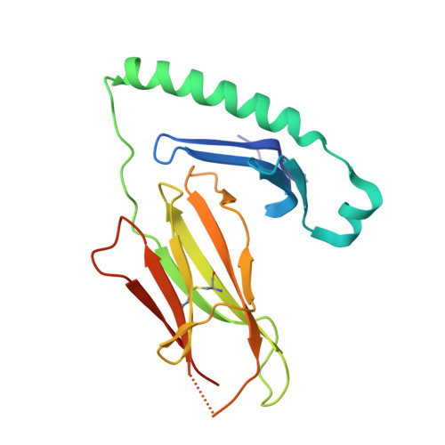
HLA class II histocompatibility antigen, DQ alpha 1 chain
A, C
192Homo sapiensMutation(s): 0 
Gene Names: HLA-DQA1
UniProt & NIH Common Fund Data Resources
Find proteins for P01909 (Homo sapiens)
Explore P01909 
Go to UniProtKB:  P01909
PHAROS:  P01909
GTEx:  ENSG00000196735 
Entity Groups  
Sequence Clusters30% Identity50% Identity70% Identity90% Identity95% Identity100% Identity
UniProt GroupP01909
Glycosylation
Glycosylation Sites: 1Go to GlyGen: P01909-1
Sequence Annotations
Expand
  • Reference Sequence
Find similar proteins by:  (by identity cutoff)  |  3D Structure
Entity ID: 2
MoleculeChains Sequence LengthOrganismDetailsImage
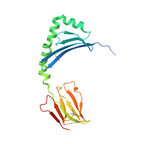
HLA class II histocompatibility antigen, DQ beta 1 chain
B, D
215Homo sapiensMutation(s): 0 
Gene Names: HLA-DQBHLA-DQB1
UniProt & NIH Common Fund Data Resources
Find proteins for P01920 (Homo sapiens)
Explore P01920 
Go to UniProtKB:  P01920
PHAROS:  P01920
Entity Groups  
Sequence Clusters30% Identity50% Identity70% Identity90% Identity95% Identity100% Identity
UniProt GroupP01920
Sequence Annotations
Expand
  • Reference Sequence
Find similar proteins by:  (by identity cutoff)  |  3D Structure
Entity ID: 3
MoleculeChains Sequence LengthOrganismDetailsImage
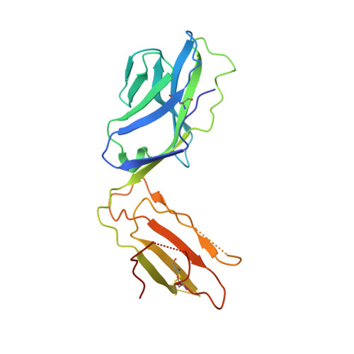
T-CELL RECEPTOR, SP3.4 ALPHA CHAIN
E, G
207Homo sapiensMutation(s): 0 
UniProt
Find proteins for K7N5N2 (Homo sapiens)
Explore K7N5N2 
Go to UniProtKB:  K7N5N2
Entity Groups  
Sequence Clusters30% Identity50% Identity70% Identity90% Identity95% Identity100% Identity
UniProt GroupK7N5N2
Sequence Annotations
Expand
  • Reference Sequence
Find similar proteins by:  (by identity cutoff)  |  3D Structure
Entity ID: 4
MoleculeChains Sequence LengthOrganismDetailsImage
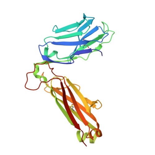
T-CELL RECEPTOR, SP3.4 BETA CHAIN
F, H
245Homo sapiensMutation(s): 0 
UniProt
Find proteins for P01850 (Homo sapiens)
Explore P01850 
Go to UniProtKB:  P01850
Entity Groups  
Sequence Clusters30% Identity50% Identity70% Identity90% Identity95% Identity100% Identity
UniProt GroupP01850
Sequence Annotations
Expand
  • Reference Sequence

Find similar proteins by:  Sequence   |   3D Structure  

Entity ID: 5
MoleculeChains Sequence LengthOrganismDetailsImage
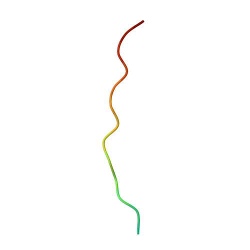
Peptide from Alpha/beta-gliadin MM1
I, J
18Triticum aestivumMutation(s): 2 
UniProt
Find proteins for P18573 (Triticum aestivum)
Explore P18573 
Go to UniProtKB:  P18573
Entity Groups  
Sequence Clusters30% Identity50% Identity70% Identity90% Identity95% Identity100% Identity
UniProt GroupP18573
Sequence Annotations
Expand
  • Reference Sequence
Experimental Data & Validation

Experimental Data

  • Method: X-RAY DIFFRACTION
  • Resolution: 3.20 Å
  • R-Value Free: 0.285 
  • R-Value Work: 0.246 
  • R-Value Observed: 0.248 
  • Space Group: P 21 21 21
Unit Cell:
Length ( Å )Angle ( ˚ )
a = 111.708α = 90
b = 134.325β = 90
c = 140.304γ = 90
Software Package:
Software NamePurpose
Blu-Icedata collection
PHASERphasing
REFMACrefinement
HKL-2000data reduction
HKL-2000data scaling

Structure Validation

View Full Validation Report


Ligand Structure Quality Assessment 


Entry History 

Deposition Data

Revision History  (Full details and data files)

  • Version 1.0: 2012-10-24
    Type: Initial release
  • Version 1.1: 2020-07-29
    Type: Remediation
    Reason: Carbohydrate remediation
    Changes: Data collection, Database references, Derived calculations, Structure summary
  • Version 1.2: 2022-08-24
    Changes: Database references, Structure summary
  • Version 1.3: 2023-11-08
    Changes: Data collection, Refinement description
  • Version 1.4: 2024-11-13
    Changes: Structure summary