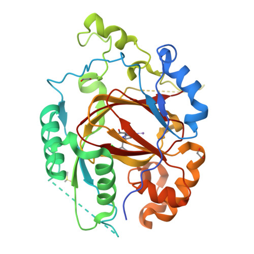
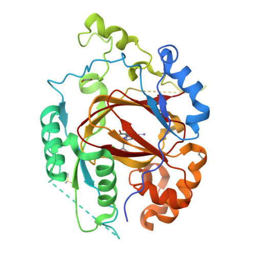
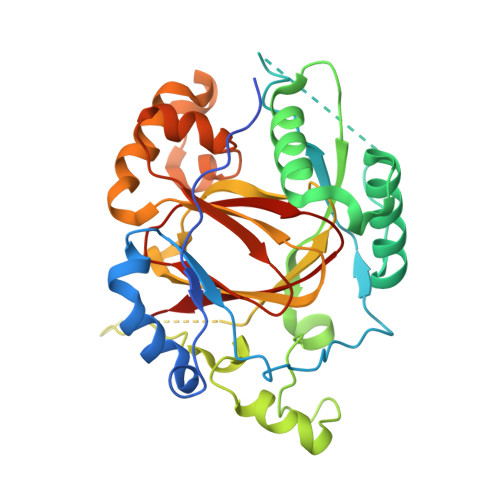
RCSB PDB - 6BH1: LINKED KDM5A JMJ DOMAIN BOUND TO THE INHIBITOR (S)-2-((2-chlorophenyl)(2-(piperidin-1-yl)ethoxy)methyl)-1l2-pyrrolo[3,2-b]pyridine-7-carboxylic acid (Compound N52)

 6BH1

LINKED KDM5A JMJ DOMAIN BOUND TO THE INHIBITOR (S)-2-((2-chlorophenyl)(2-(piperidin-1-yl)ethoxy)methyl)-1l2-pyrrolo[3,2-b]pyridine-7-carboxylic acid (Compound N52)


Experimental Data Snapshot

  • Method: X-RAY DIFFRACTION
  • Resolution: 1.93 Å
  • R-Value Free: 0.184 
  • R-Value Work: 0.160 
  • R-Value Observed: 0.161 

Starting Model: experimental
View more details

wwPDB Validation   3D Report Full Report


Ligand Structure Quality Assessment 

Created with Raphaël 2.3.0Worse 01 BetterLigand structure goodness of fit to experimental dataBest fitted DQGClick on this verticalbar to view details

This is version 1.3 of the entry. See complete history


Literature

Insights into the Action of Inhibitor Enantiomers against Histone Lysine Demethylase 5A.

Horton, J.R.Liu, X.Wu, L.Zhang, K.Shanks, J.Zhang, X.Rai, G.Mott, B.T.Jansen, D.J.Kales, S.C.Henderson, M.J.Pohida, K.Fang, Y.Hu, X.Jadhav, A.Maloney, D.J.Hall, M.D.Simeonov, A.Fu, H.Vertino, P.M.Yan, Q.Cheng, X.

(2018) J Med Chem 61: 3193-3208

  • DOI: https://doi.org/10.1021/acs.jmedchem.8b00261
  • Primary Citation of Related Structures:  
    6BGU, 6BGV, 6BGW, 6BGX, 6BGY, 6BGZ, 6BH0, 6BH1, 6BH2, 6BH3, 6BH4, 6BH5

  • PubMed Abstract: 

    Isomers of chiral drugs can exhibit marked differences in biological activities. We studied the binding and inhibitory activities of 12 compounds against KDM5A. Among them are two pairs of enantiomers representing two distinct inhibitor chemotypes, namely, ( R)- and ( S)-2-((2-chlorophenyl)(2-(piperidin-1-yl)ethoxy)methyl)-1 H-pyrrolo[3,2- b]pyridine-7-carboxylic acid (compounds N51 and N52) and ( R) - and ( S) -N-(1-(3-isopropyl-1 H-pyrazole-5-carbonyl)pyrrolidin-3-yl)cyclopropanecarboxamide (compounds N54 and N55). In vitro, the S enantiomer of the N51/N52 pair (N52) and the R enantiomer of the N54/N55 pair (N54) exhibited about 4- to 5-fold greater binding affinity. The more potent enzyme inhibition of KDM5A by the R-isoform for the cell-permeable N54/N55 pair translated to differences in growth inhibitory activity. We determined structures of the KDM5A catalytic domain in complex with all 12 inhibitors, which revealed the interactions (or lack thereof) responsible for the differences in binding affinity. These results provide insights to guide improvements in binding potency and avenues for development of cell permeable inhibitors of the KDM5 family.


  • Organizational Affiliation

    Department of Molecular and Cellular Oncology , The University of Texas MD Anderson Cancer Center , Houston , Texas 77030 , United States.


Macromolecules
Find similar proteins by:  (by identity cutoff)  |  3D Structure
Entity ID: 1
MoleculeChains Sequence LengthOrganismDetailsImage
Lysine-specific demethylase 5A, linked KDM5A JMJ domain330Homo sapiensMutation(s): 0 
Gene Names: KDM5AJARID1ARBBP2RBP2
EC: 1.14.11 (PDB Primary Data), 1.14.11.67 (UniProt)
UniProt & NIH Common Fund Data Resources
Find proteins for P29375 (Homo sapiens)
Explore P29375 
Go to UniProtKB:  P29375
PHAROS:  P29375
GTEx:  ENSG00000073614 
Entity Groups  
Sequence Clusters30% Identity50% Identity70% Identity90% Identity95% Identity100% Identity
UniProt GroupP29375
Sequence Annotations
Expand
  • Reference Sequence
Small Molecules
Ligands 4 Unique
IDChains Name / Formula / InChI Key2D Diagram3D Interactions
DQG (Subject of Investigation/LOI)
Query on DQG

Download Ideal Coordinates CCD File 
B [auth A]2-{(S)-(2-chlorophenyl)[2-(piperidin-1-yl)ethoxy]methyl}-1H-pyrrolo[3,2-b]pyridine-7-carboxylic acid
C22 H24 Cl N3 O3
AWPYWZMMJZGPOX-NRFANRHFSA-N
DMS
Query on DMS

Download Ideal Coordinates CCD File 
D [auth A]DIMETHYL SULFOXIDE
C2 H6 O S
IAZDPXIOMUYVGZ-UHFFFAOYSA-N
EDO
Query on EDO

Download Ideal Coordinates CCD File 
E [auth A],
F [auth A]
1,2-ETHANEDIOL
C2 H6 O2
LYCAIKOWRPUZTN-UHFFFAOYSA-N
MN
Query on MN

Download Ideal Coordinates CCD File 
C [auth A]MANGANESE (II) ION
Mn
WAEMQWOKJMHJLA-UHFFFAOYSA-N
Binding Affinity Annotations 
IDSourceBinding Affinity
DQG BindingDB:  6BH1 Kd: 60 (nM) from 1 assay(s)
IC50: 590 (nM) from 1 assay(s)
Experimental Data & Validation

Experimental Data

  • Method: X-RAY DIFFRACTION
  • Resolution: 1.93 Å
  • R-Value Free: 0.184 
  • R-Value Work: 0.160 
  • R-Value Observed: 0.161 
  • Space Group: C 1 2 1
Unit Cell:
Length ( Å )Angle ( ˚ )
a = 116.028α = 90
b = 61.786β = 92.47
c = 46.643γ = 90
Software Package:
Software NamePurpose
PHENIXrefinement
HKL-2000data reduction
HKL-2000data scaling
PHENIXphasing

Structure Validation

View Full Validation Report


Ligand Structure Quality Assessment 

Created with Raphaël 2.3.0Worse 01 BetterLigand structure goodness of fit to experimental dataBest fitted DQGClick on this verticalbar to view details

Entry History & Funding Information

Deposition Data


Funding OrganizationLocationGrant Number
National Institutes of Health/National Institute of General Medical Sciences (NIH/NIGMS)United StatesGM049245-23

Revision History  (Full details and data files)

  • Version 1.0: 2018-03-28
    Type: Initial release
  • Version 1.1: 2018-04-25
    Changes: Data collection, Database references
  • Version 1.2: 2020-01-01
    Changes: Author supporting evidence
  • Version 1.3: 2023-10-04
    Changes: Data collection, Database references, Derived calculations, Refinement description