6CI5 | pdb_00006ci5

Crystal structure of the formyltransferase PseJ from Anoxybacillus kamchatkensis in complex with UDP-4,6-dideoxy-4-formamido-L-AltNAc and tetrahydrofolate


Experimental Data Snapshot

  • Method: X-RAY DIFFRACTION
  • Resolution: 2.00 Å
  • R-Value Free: 
    0.240 (Depositor), 0.240 (DCC) 
  • R-Value Work: 
    0.218 (Depositor), 0.220 (DCC) 
  • R-Value Observed: 
    0.220 (Depositor) 

Starting Model: experimental
View more details

wwPDB Validation   3D Report Full Report


Ligand Structure Quality Assessment 

Created with Raphaël 2.3.0Worse 01 BetterLigand structure goodness of fit to experimental dataBest fitted F5GClick on this verticalbar to view detailsBest fitted 1YJClick on this verticalbar to view details

This is version 1.4 of the entry. See complete history


Literature

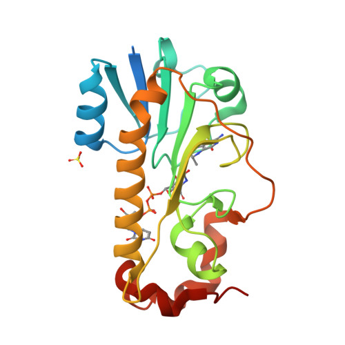
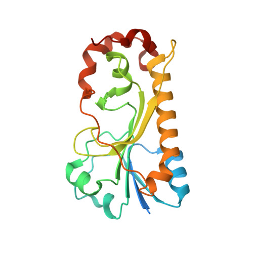
Structural Insight into a Novel Formyltransferase and Evolution to a Nonribosomal Peptide Synthetase Tailoring Domain.

Reimer, J.M.Harb, I.Ovchinnikova, O.G.Jiang, J.Whitfield, C.Schmeing, T.M.

(2018) ACS Chem Biol 13: 3161-3172

  • DOI: https://doi.org/10.1021/acschembio.8b00739
  • Primary Citation of Related Structures:  
    6CI2, 6CI4, 6CI5, 6EDK

  • PubMed Abstract: 

    Nonribosomal peptide synthetases (NRPSs) increase the chemical diversity of their products by acquiring tailoring domains. Linear gramicidin synthetase starts with a tailoring formylation (F) domain, which likely originated from a sugar formyltransferase (FT) gene. Here, we present studies on an Anoxybacillus kamchatkensis sugar FT representative of the prehorizontal gene transfer FT. Gene cluster analysis reveals that this FT acts on a UDP-sugar in a novel pathway for synthesis of a 7-formamido derivative of CMP-pseudaminic acid. We recapitulate the pathway up to and including the formylation step in vitro, experimentally demonstrating the role of the FT. We also present X-ray crystal structures of the FT alone and with ligands, which unveil contrasts with other structurally characterized sugar FTs and show close structural similarity with the F domain. The structures reveal insights into the adaptations that were needed to co-opt and evolve a sugar FT into a functional and useful NRPS domain.


  • Organizational Affiliation

    Department of Biochemistry , McGill University , Montréal , Québec H3G 0B1 , Canada.


Macromolecules
Find similar proteins by:  (by identity cutoff)  |  3D Structure
Entity ID: 1
MoleculeChains Sequence LengthOrganismDetailsImage
formyltransferase PseJ217Anoxybacillus ayderensis G10Mutation(s): 0 
Entity Groups  
Sequence Clusters30% Identity50% Identity70% Identity90% Identity95% Identity100% Identity
Sequence Annotations
Expand
  • Reference Sequence
Small Molecules
Ligands 3 Unique
IDChains Name / Formula / InChI Key2D Diagram3D Interactions
F5G
Query on F5G

Download Ideal Coordinates CCD File 
C [auth A](2R,3R,4S,5R,6S)-3-(acetylamino)-5-(formylamino)-4-hydroxy-6-methyltetrahydro-2H-pyran-2-yl [(2R,3S,4R,5R)-5-(2,4-dioxo-3,4-dihydropyrimidin-1(2H)-yl)-3,4-dihydroxytetrahydrofuran-2-yl]methyl dihydrogen diphosphate (non-preferred name)
C18 H28 N4 O16 P2
MNNFOPXYMSSSNV-XQDVVILDSA-N
1YJ
Query on 1YJ

Download Ideal Coordinates CCD File 
D [auth A]N-[4-({[(6R)-2-amino-4-oxo-3,4,5,6,7,8-hexahydropteridin-6-yl]methyl}amino)benzoyl]-L-glutamic acid
C19 H23 N7 O6
MSTNYGQPCMXVAQ-NEPJUHHUSA-N
SO4
Query on SO4

Download Ideal Coordinates CCD File 
B [auth A]SULFATE ION
O4 S
QAOWNCQODCNURD-UHFFFAOYSA-L
Experimental Data & Validation

Experimental Data

  • Method: X-RAY DIFFRACTION
  • Resolution: 2.00 Å
  • R-Value Free:  0.240 (Depositor), 0.240 (DCC) 
  • R-Value Work:  0.218 (Depositor), 0.220 (DCC) 
  • R-Value Observed: 0.220 (Depositor) 
Space Group: C 1 2 1
Unit Cell:
Length ( Å )Angle ( ˚ )
a = 96.42α = 90
b = 73.78β = 103.47
c = 41.8γ = 90
Software Package:
Software NamePurpose
phenix.refinerefinement
PHENIXrefinement
Aimlessdata scaling
DIALSdata reduction
PHENIXphasing

Structure Validation

View Full Validation Report


Ligand Structure Quality Assessment 

Created with Raphaël 2.3.0Worse 01 BetterLigand structure goodness of fit to experimental dataBest fitted F5GClick on this verticalbar to view detailsBest fitted 1YJClick on this verticalbar to view details

Entry History & Funding Information

Deposition Data


Funding OrganizationLocationGrant Number
Canadian Institutes of Health Research (CIHR)CanadaFDN-148472

Revision History  (Full details and data files)

  • Version 1.0: 2018-10-17
    Type: Initial release
  • Version 1.1: 2018-11-07
    Changes: Data collection, Database references
  • Version 1.2: 2018-11-28
    Changes: Data collection, Database references
  • Version 1.3: 2020-01-08
    Changes: Author supporting evidence
  • Version 1.4: 2023-10-04
    Changes: Data collection, Database references, Refinement description