7AE6 | pdb_00007ae6

Crystal structure of di-AMPylated HEPN(R102A) toxin


Experimental Data Snapshot

  • Method: X-RAY DIFFRACTION
  • Resolution: 1.65 Å
  • R-Value Free: 
    0.200 (Depositor), 0.200 (DCC) 
  • R-Value Work: 
    0.171 (Depositor), 0.170 (DCC) 
  • R-Value Observed: 
    0.173 (Depositor) 

Starting Model: experimental
View more details

wwPDB Validation   3D Report Full Report


Ligand Structure Quality Assessment 

Created with Raphaël 2.3.0Worse 01 BetterLigand structure goodness of fit to experimental dataBest fitted EDOClick on this verticalbar to view details

This is version 1.2 of the entry. See complete history


Literature

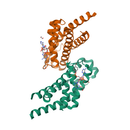
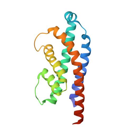
HEPN-MNT Toxin-Antitoxin System: The HEPN Ribonuclease Is Neutralized by OligoAMPylation.

Songailiene, I.Juozapaitis, J.Tamulaitiene, G.Ruksenaite, A.Sulcius, S.Sasnauskas, G.Venclovas, C.Siksnys, V.

(2020) Mol Cell 80: 955-970.e7

  • DOI: https://doi.org/10.1016/j.molcel.2020.11.034
  • Primary Citation of Related Structures:  
    7AE2, 7AE6, 7AE8, 7AE9, 7AER

  • PubMed Abstract: 

    Prokaryotic toxin-antitoxin (TA) systems are composed of a toxin capable of interfering with key cellular processes and its neutralizing antidote, the antitoxin. Here, we focus on the HEPN-MNT TA system encoded in the vicinity of a subtype I-D CRISPR-Cas system in the cyanobacterium Aphanizomenon flos-aquae. We show that HEPN acts as a toxic RNase, which cleaves off 4 nt from the 3' end in a subset of tRNAs, thereby interfering with translation. Surprisingly, we find that the MNT (minimal nucleotidyltransferase) antitoxin inhibits HEPN RNase through covalent di-AMPylation (diadenylylation) of a conserved tyrosine residue, Y109, in the active site loop. Furthermore, we present crystallographic snapshots of the di-AMPylation reaction at different stages that explain the mechanism of HEPN RNase inactivation. Finally, we propose that the HEPN-MNT system functions as a cellular ATP sensor that monitors ATP homeostasis and, at low ATP levels, releases active HEPN toxin.


  • Organizational Affiliation

    Institute of Biotechnology, Life Sciences Center, Vilnius University, Sauletekio av. 7, 10257 Vilnius, Lithuania.


Macromolecules
Find similar proteins by:  (by identity cutoff)  |  3D Structure
Entity ID: 1
MoleculeChains Sequence LengthOrganismDetailsImage
HEPN toxin
A, B
157Aphanizomenon flos-aquae 2012/KM1/D3Mutation(s): 1 
Gene Names: OA07_26455
EC: 3.1.27
UniProt
Find proteins for A0A0B0QJR1 (Aphanizomenon flos-aquae (strain 2012/KM1/D3))
Explore A0A0B0QJR1 
Go to UniProtKB:  A0A0B0QJR1
Entity Groups  
Sequence Clusters30% Identity50% Identity70% Identity90% Identity95% Identity100% Identity
UniProt GroupA0A0B0QJR1
Sequence Annotations
Expand
  • Reference Sequence
Small Molecules
Ligands 2 Unique
IDChains Name / Formula / InChI Key2D Diagram3D Interactions
AMP
Query on AMP

Download Ideal Coordinates CCD File 
C [auth A],
D [auth A],
H [auth B],
I [auth B]
ADENOSINE MONOPHOSPHATE
C10 H14 N5 O7 P
UDMBCSSLTHHNCD-KQYNXXCUSA-N
EDO (Subject of Investigation/LOI)
Query on EDO

Download Ideal Coordinates CCD File 
E [auth A]
F [auth A]
G [auth A]
J [auth B]
K [auth B]
1,2-ETHANEDIOL
C2 H6 O2
LYCAIKOWRPUZTN-UHFFFAOYSA-N
Experimental Data & Validation

Experimental Data

  • Method: X-RAY DIFFRACTION
  • Resolution: 1.65 Å
  • R-Value Free:  0.200 (Depositor), 0.200 (DCC) 
  • R-Value Work:  0.171 (Depositor), 0.170 (DCC) 
  • R-Value Observed: 0.173 (Depositor) 
Space Group: P 21 21 21
Unit Cell:
Length ( Å )Angle ( ˚ )
a = 60.443α = 90
b = 62.722β = 90
c = 84.356γ = 90
Software Package:
Software NamePurpose
PHENIXrefinement
XDSdata reduction
SCALAdata scaling
MOLREPphasing
PDB_EXTRACTdata extraction

Structure Validation

View Full Validation Report


Ligand Structure Quality Assessment 

Created with Raphaël 2.3.0Worse 01 BetterLigand structure goodness of fit to experimental dataBest fitted EDOClick on this verticalbar to view details

Entry History 

Deposition Data

Revision History  (Full details and data files)

  • Version 1.0: 2020-12-30
    Type: Initial release
  • Version 1.1: 2024-01-31
    Changes: Data collection, Database references, Refinement description
  • Version 1.2: 2024-11-06
    Changes: Structure summary